
Institut Charles Delaunay-Universite de technologie de Troyes, CNRS FRE 2848, Laboratoire de Nanotechnologie et d'InstrumentationOptique, 12 rue Marie Curie, BP 2060 - 10010 Troyes Cedex, France
E.mail : adam@utt.fr
Keywords: plasmonics, biosensors, surface enhanced Raman scattering, metal enhanced fluorecence
Plasmonics is now well established field finding numerous applications in pharmacology, biology, photonic circuitry and metamaterials among others. For the detection of hazardous molecules or markers Surface Enhanced Spectroscopies are well widespread [1]. Among them, Surface Enhanced Raman Spectroscopy (SERS) and Metal Enhanced Fluorescence (MEF) are the most used for applications. Both enhanced spectroscopies are based on local field enhancement entailed in the near vicinity of metallic nanoparticles when Surface Plasmon oscillations are driven for a specific optical wavelength. Another approach for detecting various molecules is biosensors relying on the detection of the spectral shift of the Surface Plasmon resonance of metallic nanoparticles after the adsorption of these same molecules.
In this contribution it will be presented what plasmonics can bring to surface enhanced spectroscopies and show the ties between SERS and MEF. In that direction some of the works of the LNIO laboratory will be presented [2-3].
[1] K. M. Kosuda, J. M. Bingham, K. L. Wustholz, and R. P. Van Duyne, Chapter 110, In Handbook of Nanoscale Optics and Electronics; Wiederrecht, G., Ed., Elsevier, Amsterdam (2010).
[2] J. Grand, J., M. Lamy de la Chapelle, J.L. Bijeon, P.M. Adam , A. Vial, P. Royer , Phys. Rev. B, 72, 033407 (2005)
[3] P. Viste, J. Plain, R. Jaffiol, A. Vial, P.-M. Adam, P. Royer, ACS Nano, 4, 759 (2010)
Department of Electronic Engineering, Ching Yun University, Jung-Li, Taiwan 320, R.O.C.
We numerically report on first realization of near field interaction and localized surface plasmon resonance of a pair of silver-shell nanospheres with different dielectric holes embedded in a dielectric substrate using finite element method. An electromagnetic mode different from the solid case of the same volume is excited inside and outside the shell surface, resulting in an intensity enhancement in a gap of particle pair surrounding the particle-substrate interface. We find that the embedded depth of the nanoparticles pair in a substrate will influence the position of the localized fields which is confined in the gap. Besides, the near field intensity becomes less intense and the spectrum of peak resonances red-shifted as the index difference of interface and embedded depth decreases. The proposed models have been prepared to cover the visible and NIR regions with application to sensors and spectroscopy purposes by varying the embedded depth and pertinent to the functionality of sensors, spectroscopy and other optical devices.
The authors acknowledge the financial support from the National Science Council of the Republic of China (Taiwan) under Contract No. NSC 99-2112-M-231-001-MY3 and NSC-99-2120-M-002-12.
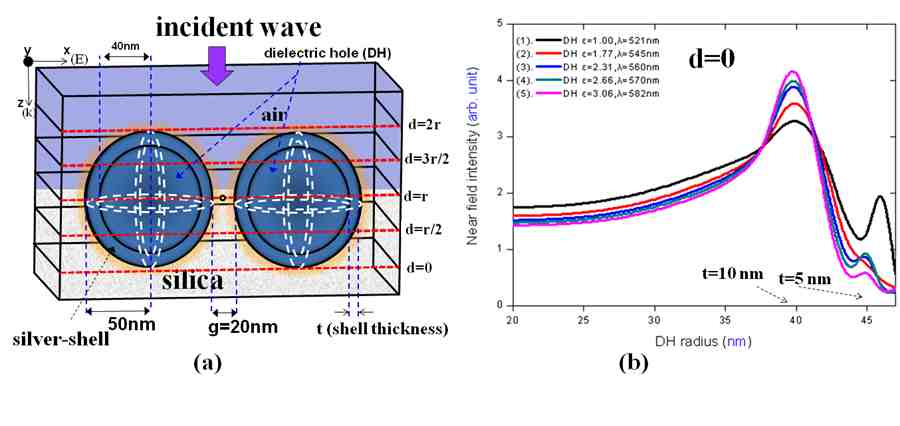
- Fig. 1: (a) Schematic of two identical silver-shell nanospheres with shell thickness t embedded in a silica substrate, (b) Near field intensities of DH case (d=0) with different dielectric medium in DHs corresponding to their SPR peak wavelengths as a function of DH raidus (inner radius of nonospheres).
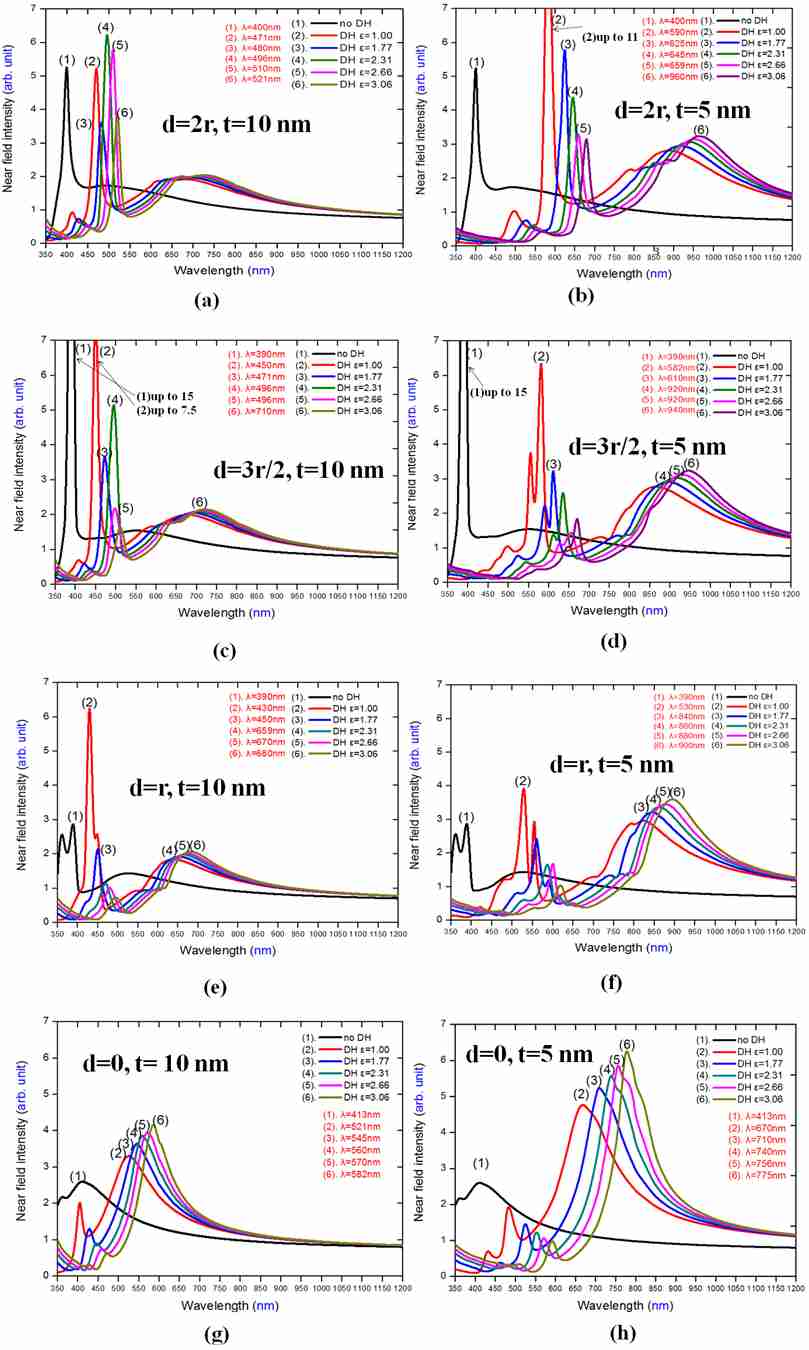
- Fig. 2: Compare near field intensities of no DH case (d=2r) and DH cases [(d=2r, t=10 nm) and (d=2r, t=5 nm) respectively] embedded in a silica substrate as a function of wavelengths.
[1] Y.-F. Chau, H-H Yeh and D. P. Tsai, Appl. Opt. Vol. 47, 5557-5561, 2008.
[2] Y.-F. Chau, Plasmonics, 4: 253-259 (2009).
[3]. Nehl, C. L., Liao, H. and Hafner, J. H., Nano Lett. Vol. 6, 683-688, 2006.
[4]. Y.-F. Chau, H.-H. Yeh and D. P. Tsai, J. of Electromagn. Waves and appli., Vol. 24, 1005-1014, 2010.
Institute of Applied Physics, Friedrich Schiller University Jena, Max-Wien Platz 1, D-07743 Jena, Germany
Metamaterials are composites consisting of artificial metaatoms/metamolecules with typical sizes less than the respective wavelengths. A key point of the metamaterials, which differs them principally from the natural one (at least in a visible wavelength range) is the magnetic response on an external electric field. In this work we summarize the multipole expansion approach in order to describe analytically linear and nonlinear effective optical properties of passive metamaterials. In order to validate our model, we applied the formalism for the split-ring resonator and the cut-wire structure and compare the results with rigorous numerical calculations. A new type of the second order nonlinearity - so called multipole nonlinearity - has been found to appear even for intrinsically linear metal, which the nano resonators consist of. We also report about extension of the model in order to describe optical properties of metamaterials coupled with various quantum systems, namely Quantum Dots (QD) and Carbon Nano Tubes (CNT). The regular dynamics (transient dynamics and stabilized generation) and stochastic properties (linewidth of generation) of a nano laser - metamaterial coupled with QD - have been described using the proposed model and compared with the published experimental data. By pumping of the appropriately positioned Quantum Dots (QDs) as a gain media, a magnetic response without optical losses can be obtained. Nonlinear CNT properties modification due to coupling with resonant metamaterials has been compared with respective experimental data.
Institute of Photonic Technology (IPHT) Jena, Germany
Microstructured optical fibers (MOFs) and especially suspended core fibers with small (micrometer) core diameter in combination with plasmonic nanoparticles represent a large potential for novel sensor devices.
Plasmonic nanoparticles can be designed and synthesized with different materials, dimensions and shapes by cost-effective chemical ways. The plasmonic properties of such particles are due to the collective oscillation of the conductive electrons and result in the existence of the localized surface plasmon resonance (LSPR) of particle solutions or the strong scattering properties at the single particles. The position of the LSPR band can be tuned from the UV to the near infrared spectral range. The newly developed nanoparticle layer deposition (NLD) technique coats the inner walls of MOFs or SCFs with selected plasmonic particles. The NLD method is based on self-assembly monolayer techniques and on microfluidics and induces a particle deposition over several meter lengths with homogeneous coating density. The resulting plasmonically tuned fibers show the same optical properties as the selected particle type. In the poster we will present examples for the sensing properties of such plasmonically tuned optical fibers for bioanalytics, e.g. for the DNA-based detection of phytopathogenic microorganisms.
Laboratoire de Nanotechnologie et d'Instrumentation Optique LNIO-ICD, UTT, France
A. Bouhelier
Laboratoire Interdisciplinaire Carnot de Bourgogne CNRS-UMR 5209, UB, France
P. K. Jain
Miller Institute for Basic Research in Science and Department of Chemistry, UCB, USA
O. Soppera
Institut de Science des Materiaux de Mulhouse IS2M-CNRS LCR 7228, UHA, France
The nature of near-fields around optical nanostructures has been a topic of great fundamental and practical interest. The near-field properties of metal nanostructures are typically accessible only by optically exciting the nanostructure in resonance with its plasmon mode/s [1,2]. A lot has been learnt about the plasmon modes and associated near-fields of the nanostructure [3]; however, one of the most basic questions remains unanswered: What is the fundamental difference between the optical characteristics of a nanostructure illuminated off its resonance from those under resonant excitation of one of the modes of the nanostructure? So far it has been difficult to approach this question due to the difficulty of imaging nanostructures spectrally away from their strong plasmon resonance modes.
In this work, we report on the imaging of a metallic nanostructure away from its resonance. After morphological characterization using atomic force microscopy (AFM), the gold nanorods are coated with a photopolymerizable formulation and illuminated out of their resonance, at a wavelength λ = 530 nm that matches the maximum of the photopolymerizable formulation absorption spectrum, but sufficiently off-resonant from the nanorod surface plasmon responses.
We realize that the laser illumination of the nanostructure induces surface charges at the metal/dielectric interface, resulting in a local enhancement of the electric field normal to the interface [2]. Away from the plasmon resonance, this field enhancement is considerably weak spectrally. However, it is large enough to be exploited for initiating the local photopolymerization of a monomeric system, thus providing a "molecular mold" of the photoinduced surface charge distribution around the nanostructure, as illustrated in Fig. 1. This photochemical approach has already been employed to image the resonant behavior of metallic nanoparticles (i.e. their surface plasmon resonance) [2].
We show here that the sensitivity of the photopolymer is high enough to imprint the local non-resonant field with nanoscale resolution, i.e. sub-5-nm, thus allowing what we believe to be the first visualization of the surface charge density distribution signature [4]. We are also able to deduce from our measurements, the real part of the dielectric function of the metal that comprises the nanostructure [4].
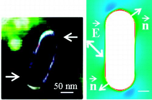
- Fig. 1: Differential image highlighting the very localized polymerization that occurs at the surface of the gold nanorod, together with the FDTD simulations.
[1] W. L. Barnes, A. Dereux, and T. W. Ebbesen. Nature 424, 824-830 (2003).
[2] C. Deeb, R. Bachelot, J. Plain, A. L. Baudrion, S. Jradi, A. Bouhelier, O. Soppera, P. K. Jain, L. Huang, C. Ecoffet, L. Balan, and P. Royer. ACS Nano 4, 4579-4586 (2010).
[3] S. A. Maier. Plasmonics, Springer (2007).
[4] C. Deeb, X. Zhou, D. Gerard, A. Bouhelier, P. K. Jain, J. Plain, O. Soppera, P. Royer, and Renaud Bachelot. J. Phys. Chem. Lett. 2, 7-11 (2011).
1Laboratory for Theoretical Physics, University Paul Sabatier, CNRS, Toulouse, F-31062, France
2Institute for Theoretical Physics, University of Erlangen-Nuremberg, Erlangen, G-91058, Germany
Metal clusters are known to exhibit collectives modes, namely surface plasmon resonances, which can express as sharp peaks in the optical response. In the framework of time-dependent density functional theory with real-time propagation in real space, we will review in this talk various ways to exploit the Mie plasmon resonance of small sodium clusters, via their optical response, in dynamical scenarios: pump and probe measurements in cluster fission [1], Coulomb explosion of clusters embedded in a rare gas matrix [2,3,4], chromophore effect in a water molecule.
[1] P.M. Dinh, P.-G. Reinhard and E.Suraud, Time resolved fission in metal clusters, J. Phys. B 38, 1637 (2005)
[2] F. Fehrer, P.M. Dinh, P.-G. Reinhard, and E. Suraud, Embedded metal cluster in strong laser fields,
Comp. Mat. Sci. 42 (2008) 203
[3] F. Fehrer, P.M. Dinh, M. Baer, P.-G. Reinhard, and E. Suraud, Hindered Coulomb explosion of embedded Na clusters - stopping, shape dynamics and energy transport, Euro. Phys. J. D 45 (2007) 447
[4] P. M. Dinh, P.-G. Reinhard, E. Suraud, Dynamics of clusters and molecules in contact with an environment, Phys. Rep. 485 (2010) 43
1Laboratoire CSPBAT, Université Paris VIII, 93017 Bobigny, France.
2Institut Fresnel, Aix-Marseille Université, CNRS, Ecole Centrale Marseille, Campus de St Jérôme, 13397 Marseille, France.
3Institut de Science et d'Ingénierie Supramoléculaires, Université de Strasbourg, CNRS, 8 allée G. Monge, 67000 Strasbourg, France.
Arrays of nanoapertures have been demonstrated to realize efficient, robust, and reproducible substrates for surface-enhanced Raman scattering SERS spectroscopy(1-3). However, little attention has been devoted to single nanoapertures, although a thorough understanding of the SERS phenomenon in a single aperture is essential for the rationale optimization of nanoaperture arrays for SERS(4). Single nanoapertures milled in optically thick gold films are quantitatively evaluated for the first time to determine the SERS enhancement factors using para-mercaptoaniline as nonresonant analyte. We determine a peak enhancement factor of 2×10⁵ for a single 100 nm diameter aperture (5). Nanoapertures will be functionnalized by standard avidin–biotin chemistry. The highly specific interaction of avidin with biotin (vitamin H) is mostly used in designing biosensors because of the high affinity of avidin for biotin as the non-covalent interaction of a protein and ligand. First, SERS spectra in nanoholes of avidin-biotin will be showed and finally, we will consider the possibility of transferring this process to proteins/antibody systems such as BSA/ Anti-BSA. SERS enhancement factors will be determined for each diameter of nanohole.
1. Wenger, J., Gerard, D., Lenne, P. F., Rigneault, H., Dintinger, J., Ebbesen, T. W., Boned, A., Conchonaud, F., and Marguet, D. (2006) Dual-color fluorescence cross-correlation spectroscopy in a single nanoaperture: towards rapid multicomponent screening at high concentrations. Optics Express 14, 12206-12216.
2. Wenger, J., Cluzel, B., Dintinger, J., Bonod, N., Fehrembach, A. L., Popov, E., Lenne, P. F., Ebbesen, T. W., and Rigneault, H. (2007) Radiative and nonradiative photokinetics alteration inside a single metallic nanometric aperture. Journal of Physical Chemistry C 111, 11469-11474.
3. Wenger, J., Conchonaud, F., Dintinger, J., Wawrezinieck, L., Ebbesen, T. W., Rigneault, H., Marguet, D., and Lenne, P. F. (2007) Diffusion analysis within single nanometric apertures reveals the ultrafine cell membrane organization. Biophysical Journal 92, 913-919.
4. Wenger, J., Dintinger, J., Bonod, N., Popov, E., Lenne, P. F., Ebbesen, T. W., and Rigneault, H. (2006) Raman scattering and fluorescence emission in a single nanoaperture: Optimizing the local intensity enhancement. Optics Communications 267, 224-228.
5. Djaker, N., Hostein, R., Devaux, E., Ebbesen, T. W., Rigneault, H., and Wenger, J. (2010) Surface Enhanced Raman Scattering on a Single Nanometric Aperture. Journal of Physical Chemistry C 114, 16250-16256.
Department of Bioelectronics, Institute of Solid State Electronics, TU Vienna, Vienna, Austria, dodt@tuwien.ac.at
It would be very helpful for the analysis of neuronal networks of the brain, if one could visualize these networks in 3 dimensions. Up to now this was only possible with limited resolution by sequential slicing and reconstruction of the brain. This time consuming attempt is easily hampered by artifacts as shrinkage and distortion induced by standard histological procedures.
To overcome these problems we used a microscopy based on extreme darkfield illumination with a light sheet, once called ultramicroscopy. This microscopy allows optical sectioning of whole mouse brains and was combined with an approach to clear fixed neuronal tissue: Mouse brains were made completely transparent by immersion in oil of the same refractive index as protein. By illuminating the brains with blue light (λ= 488 nm), neurons labeled with GFP were visualized by fluorescence. This way we could detect single neurons in hippocampi inside whole brains.
By surface rendering the shape and position of hippocampi relative to the brain surface could be depicted. In complete excised hippocampi subcellular resolution was obtained by 3D reconstruction from several hundred optical sections. The dendritic trees of CA1 hippocampal neurons with dendrites and dendritic spines could be visualized.
Many proteins can be labelled in transgenic mice with genetically encoded fluorescent markers. Using these markers our approach will represent a high-throughput screening method for protein expression in 3 D. This expression can be monitored with µm resolution and should allow the elucidation of complex neuronal networks in the brain.
In a genome-wide RNAi screen, flight defective Drosophila were isolated. A number of these lines show strong flight muscle defects, suggesting a muscle formation defect or extensive muscle degeneration. In our ongoing study we investigate the morphology of individual muscles in the intact fly. This enables us to assign the observed defects to particular muscle classes.
We show that ultramicroscopy allows also optical sectioning and detailed 3D reconstruction of whole mouse embryos by imaging autofluorescent structures. Especially the circulatory system in the body and brain became apparent as blood remaining in the preparation showed strong fluorescence. Also other applications like e.g. visualization of nerve bundles in whole embryos and visualization of plaques in brains of mice with Alzheimers disease will be shown. In general the method is well suited for high-throughput phenotype screening of transgenic mice and thus will benefit the investigation of disease models.
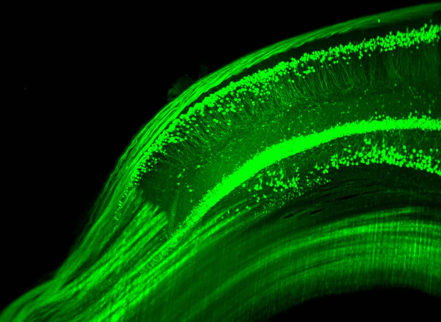
- GFP labelled hippocampus imaged with ultramicroscopy
1)BAM Federal Institute for Materials Research and Testing, Richard-Willstätter-Str. 11, 12489 Berlin
2)Humboldt-Universität zu Berlin, Department of Chemistry, Brook-Taylor-Str. 2, 12489 Berlin
3)present address: PTB Physikalisch-Technische Bundesanstalt, Magnusstraße 9, 12489 Berlin
E-Mail: daniela.drescher@bam.de
The cellular uptake of gold or silver nanoparticles allows spatially resolved analysis of cellular compartments in vitro and has proven surface-enhanced Raman scattering (SERS) to be a very useful method for the investigation of biological processes in cellular systems. Distribution and biocompatibility of SERS nanoprobes composed of gold nanoparticles and non-fluorescent Raman labels as reporter molecules will be shown. The concept of SERS hybrid probes enables the sensitive detection of intrinsic cellular constituents beside extrinsic molecules introduced into the cells for monitoring physiological parameters like pH value[1] or for tracking their biodistribution. These nanoprobes were used for Raman chemical imaging of live cells exploiting their multifunctional and multiplexing capabilities by simultaneous incubation of multiple probes. For the discrimination of SERS labels and nanoprobes, multivariate statistics were applied.[2] The results of cluster methods and principal components approaches for discrimination indicate that fast, multivariate evaluation of whole sets of multiple probes is feasible, beyond the visual inspection of individual spectra that has been practiced so far. Cytotoxicity studies indicate that our SERS nanoprobes are not toxic regarding cell morphology and metabolic activity after 24h exposure to 3T3 fibroblast cells. Especially for in vivo applications, biocompatibility is an essential property of a nanoparticulate SERS probe.
[1] J. Kneipp, H. Kneipp, B. Wittig, and K. Kneipp, J. Phys. Chem. C 2010, 114, 7421–7426,
[2] A. Matschulat, D. Drescher, J. Kneipp, ACS Nano 2010, 4, 3259-3269.
Pharmadiagnostics NV, Z1 Research Park 310, B-1731 Zellik, Belgium.
A few decades ago, it became clear that, because of their half-life and negative environmental issues, radioisotopes had to be replaced by other labels in immuno- and receptor assays. Enzymes and fluorophores became more and more popular but assay formats were still heterogeneous, i.e. requiring the separation of bound from free before reading the label signal. Another and parallel trend in the late 80s and early 90s saw the progressive advent of "label-free" surface plasmon resonance (SPR) biosensors. SPR spectroscopy allowed not only for homogeneity in assay design but also to monitor the biomolecular interactions in real time. In this context, molecular plasmonics with noble metal nanostructures became rapidly a possible interesting alternative to both labeled assays and label-free biosensors. At the time, Leuvering's group at Organon had developed the first clinical immunoassay systems based on optical detection of plasmon coupling. They used gold nanospheres conjugated with polyclonal antibodies reacting in solution with the antigen, of which specific aggregation could be detected and quantified by the large red-shift of the plasmon resonance absorbance wavelength. Using the same optical principle, Chad Mirkin's group published DNA hybridization assays. These assays were homogeneous (i.e. mix and read) and did not require any special spectroscopic instrumentation other than a photometer, or even the naked eye.
The effect of refractive index changes in the medium by chemical means on the localized SPR (LSPR) light absorbance of noble metal nanostructures was known for quite a long time. In 1998, I published an article (Analyst, 123, 1599) showing that ligand-antibody interactions induced changes in the dielectric at gold nanosphere surfaces that could be used to monitor spectroscopically and in real-time the resulting changes in the plasmon-polariton dipole resonance. Our group extended in 2001 the demonstration of this effect to low molecular weight molecules-protein binding interactions in buffered solution. The possibilities of this new application of molecular plasmonics in life sciences attracted the interest of many groups. The first applications were in diagnostics with the development of immunoassays for small or polypeptide molecules and in biotechnology with real-time monitoring of kinetics of ligand-protein interactions. Many of the early proof of concept studies attempted to mimic SPR biosensors. Nanoparticles were attached to a solid substrate and conjugated to proteins. Biomolecular interactions were then monitored in a spectrophotometer.
I will review this historical evolution of the application of molecular plasmonics in life sciences with a particular emphasis on the more recent advances in diagnostics and biotechnology. These recent advances include the development of better performing nanomaterials in terms of photonic reactivity and sensitivity, or allowing to multiplex reactions. Composite nanomaterials will also be covered which allow for new applications in drug discovery, including physico-chemical characterization, early in vitro pharmacokinetic evaluation of new chemical entities and the assessment of their interactions with pharmacological targets. The presentation will conclude with future applications of this technology in cell-based assays.
Babes-Bolyai University, Faculty of Physics and Interdisciplinary Research Institute on Bio-Nano-Sciences Nanobiophotonics Center,T. Laurian 42, 400271 Cluj-Napoca, Romania
There is an increasing interest in anisotropic noble-metal nanoparticles, particularly gold nanorods (GNRs), due to their large and controllable tunability of the surface plasmon resonances, recognized biocompatibility and versatility to incorporate multiple functionalities [1,2]. In our laboratory we currently prepare GNRs of aspect ratio ranging from 2.6 to 4.1 by cetyltrimethylammonium bromide (CTAB)-assisted chemical method. We characterize their properties by UV-Visible spectroscopy, transmission/scanning electron microscopy (SEM/TEM), dynamic light scattering (DLS) and Finite-Difference Time-Domain (FDTD). Recently we achieved to demonstrate their potential to operate as dual plasmonic amplifiers in surface-enhanced Raman scattering and metal-enhanced fluorescence (MEF).
In view of future biomedical applications it is imperative to reduce the inherent toxicity exhibited by CTAB coating. Therefore, we succeeded to encapsulate the GNRs in a uniform silica shell. Silica coating endows the GNRs with chemical stability, biocompatibility and rich surface chemistry and high porosity for further biofunctionalization [3]. The silica shell enables the GNRs conjugation with different types of biomolecules thus obtaining promising optical probes suitable for investigation in biological media. As for example we successfully recorded the surface-enhanced Raman scattering and fluorescence signals independently generated by the obtained hybrid nanostructures doped with Raman reporters and fluorescent molecules. Silica-coated gold nanorods offer multiple functionalities for biomedical applications including SERS, metal-enhanced fluorescence, photothermal therapy, intracellular detection and imaging.
This work was supported by CNCSIS in the frame of the PN-II research programs under the project PCCE No 129/2008. Ana- Maria Gabudean also acknowledges Ph.D. scholarship, project co-financed by the Sectoral Operational Program for Human Resources Development 2007-2013, Contract no. POSDRU/88/1.5/S/60185 – "Innovative doctoral studies in a knowledge based society", Babes-Bolyai University, Cluj-Napoca, Romania
[1] S. Boca, D. Rugina, A. Pintea, L. Barbu-Tudoran, S. Astilean, Nanotechnology, 2011, 22, 055702
[2] F. Cai, J. Qian, L. Jiang, S. He, J Biomed Opt. 2011, 16, 016002
[3] Q. Zhan, J. Qian, X. Li, S. He, Nanotechnology, 2010, 21, 055704
Institut Charles Delaunay, Universite de Technologie de Troyes, Laboratoire de Nanotechnologie et d'Instrumentation Optique, UMR CNRS STMR 6279, 12 rue Marie Curie BP 2060, 10010 Troyes cedex, France
Due to the high sensitivity of the localized surface plasmon resonance of metallic nanoparticles (MNPs) to the local refractive index[1], MNPs can be used as very sensitive and selective markers for the detection of molecules in very low concentration. Consequently, the last decade has seen a growing development of plasmonic nanosensors for the detection of chemical and biochemical[2] agents or for medical diagnosis[3]. MNPs have thereby a tremendous interest for both fundamental physics and biochemistry and subsequent technological applications.
In this context, we report on the achievement, characterization and application of very innovative hybrid plasmonic nanoantenna. Our approach relies on the structuration of glass substrate under the shape of glass microtips which apexes are chemically functionalized with one to ten 40 nm diameter gold colloidal nanoparticles. A SEM image of functionalized microtips is shown on figure 1. Glass microtips are acting as microaxicons, optimizing both excitation and the collection of the nanoparticle LSPR at their apexes. We demonstrate that the LSPR signal can be measured by mean of simple extinction spectroscopy setup on a single microtip functionalized with a low number of localized gold MNPs, down to single one as it can be seen on figure 2. FDTD simulations have been performed to emphasize this assumption by calculating the electric field at the gold functionalized glass microtip apex as it is depicted in figure 3. The microlense effect couples the focused photonic beam going out from the glass tip to the LSPR of the MNP redirecting the light in far field, thus acting as an antenna.
Finally, combining extinction spectrum measurements with SEM characterization, optical and morphological properties are correlated for a set of individual gold functionalized microtips. We demonstrate that a single gold functionalized microtip can be used as an isolated plasmonic nanosensor for the detection of molecules with a concentration lower than 10-12 M by extinction spectroscopy. Besides, Surface Enhanced Raman Spectroscopy properties of those nanosensors are investigated.
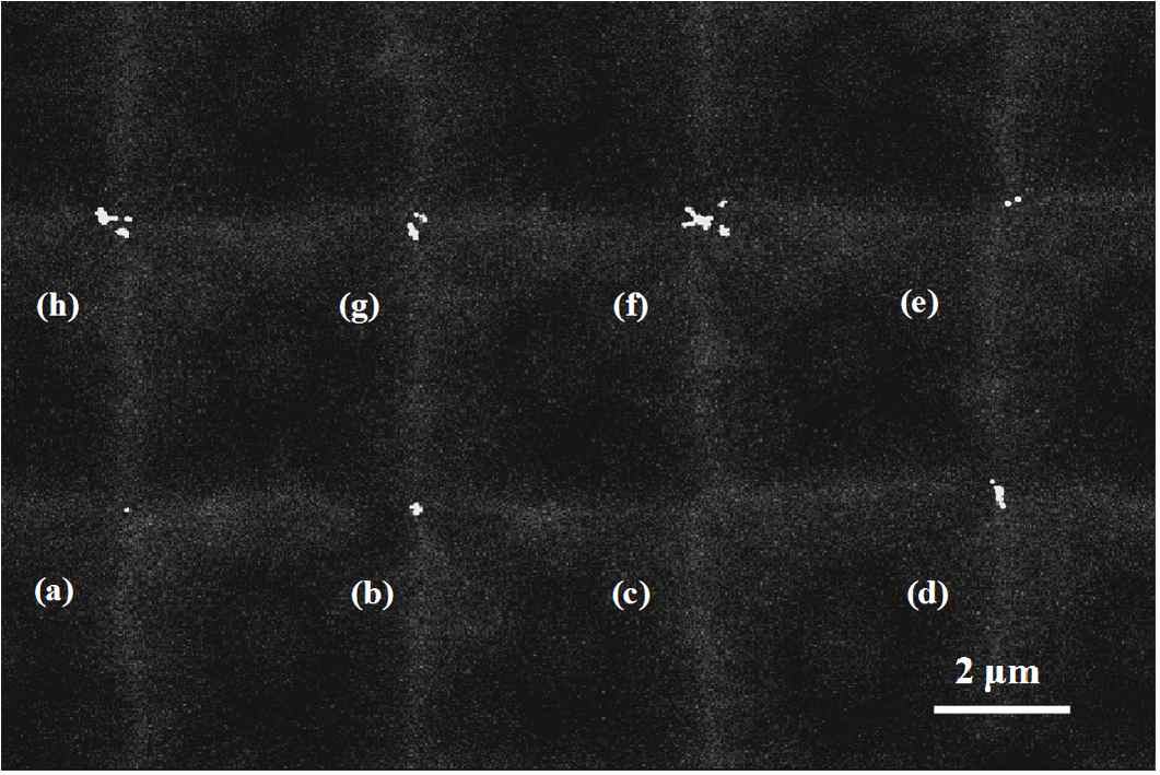
- Fig. 1: SEM image of arrays of gold functionalized glass microtips.
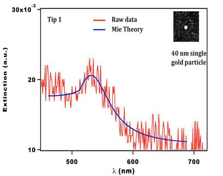
- Fig. 2: Extinction spectrum of a single gold nanoparticle.
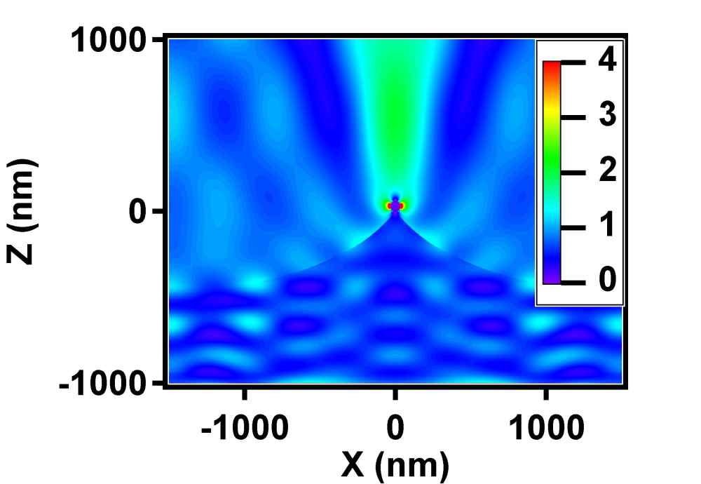
- Fig. 3: Map of the electric field at the glass apex functionalized with a single gold MNP.
[1] T. R. Jensen, M. L. Duval, L. Kelly, A. Lazarides, G. C. Schatz, R. P. Van Duyne, J. Phys. Chem. B 103, 9846-9853 (1999).
[2] A. D. McFarland and R. P. Van Duyne, Nano Lett. 3, 1057-1062 (2003).
[3] C. Loo, A. Lin A, L. Hirsch , M. Lee, J. Barton , N. Halas, J. West, R. Drezek R, Technol. Cancer Res. Treat. 3, 33-40 (2004).
Ecole Polytechnique Federale de Lausanne (EPFL), Optics Laboratory, 1015 Lausanne, Switzerland Friedrich Schiller University Jena, Institute of Applied Physics, Multiphoton Lab, 07745 Jena, Germany
Nonlinear optical processes such as second-harmonic generation (SHG) are generally inefficient at very small scale. As shown for silica-core Au nanoshells [1], it is possible to strongly enhanced the SHG response of non centrosymmetric nanoparticles by developing nonlinear optical plasmonics core shell cavities [2]. In theory, the enhancement can reach 3 orders of magnitude based on a 100 nm diameter BaTiO3 core and a 10 nm Au shell. This core-shell structure allows for reaching a near infrared plasmonics resonance and enhancing the SHG signal from nanostructures that can be used as imaging probes or localized light source in the visible spectrum range. Indeed, These nanomaterials possess several key advantages, compared to conventional fluorescent markers such as organic dyes, green fluorescent proteins (GFPs) or quantum dots, including long-term observation without photobleaching, flexibility in excitation wavelength, coherent signals for three-dimensional imaging, narrow signal bandwidth for greater noise rejection, ultrafast response time, and excellent biocompatibility.
The gold coating process involved three main steps. First of all, primary amines are coated on the surface of the bare nanoparticles [3]. The amino complex presents NH2 complex at the surface of the nanoparticle. Then, 2-3 nm gold particles are adsorbed onto the surface (Fig. 1 a). Finally, the Au shell is grown all around the particle from the seeded particles by a reaction of reduction of gold by hydroxylamine (Fig.1 b-d).
Fig. 1 (e) shows the measured extinction spectrum of the BaTiO3/Au nanocavities. The plasmonic resonance peak is in the near infrared wavelength centered around 780 nm. Because of the large size variation of the core, the resonance shows a wide inhomogeneous broadening. With this structure, enhancement of the SHG signal will be shown.
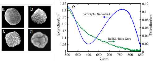
- Fig.1. (a-d) SEM pictures of different steps of the 10-nm gold shell synthesis around 100 nm (diameter) BaTiO3 nanoparticles, (e) Measured ensemble extinction spectrum of BaTiO3/Au core-shell nanocavities (blue curve) and the bare BaTiO3 nanocrystals (green curve).
[1] S. J. Oldenburg, R. D. Averitt, S. L. Westcott, and N. J. Halas, "Nanoengineering of optical resonances," Chemical Physics Letters, vol. 288, pp. 243-247, 1998.
[2] Y. Pu, R. Grange, C.-L. Hsieh, and D. Psaltis, "Nonlinear optical properties of core-shell nanocavities for enhanced second-harmonic generation," Physical Review Letters, vol. 104, pp. 207401-207405, 2010.
[3] C.-L. Hsieh, R. Grange, Y. Pu, and D. Psaltis, "Three-dimensional harmonic holographic microcopy using nanoparticles as probes for cell imaging," Opt. Express, vol. 17, pp. 2880-2891, 2009.
Marina Gühlke, Humboldt-Universität zu Berlin, Institut für Chemie, Brook-Taylor-Str. 2, 12489 Berlin
Surface-enhanced Raman-scattering (SERS) has been established as a useful tool for analytical and bioanalytical applications. SERS results in an increase of the intensity of Raman signals by several orders of magnitude due to the presence of plasmonic nanostructures.
For this purpose, mostly gold- or silver-nanoparticles, either in colloidal suspension or immobilized on solid supports, are used. Especially combinations of magnetic materials, e.g. iron oxides, and silver or gold would be attractive for analytical applications as they exhibit the plasmonic properties which are necessary for SERS, as well as magnetic properties. This kind of nanoparticles can be used for specific separation or enrichment of proteins,[1] cells[2] or other analytes with a subsequent detection by SERS.[3]
In our work combined nanostructures of iron(III) oxide and silver have been synthesized by reducing silver in the presence of iron oxide nanoparticles. The resulting structures exhibit plasmonic properties similar to those of silver nanoparticles and their SERS-activity could be demonstrated with different analytes. Furthermore the magnetic properties have been utilized in a separation experiment to examine adsorption of analyte molecules in SERS-measurements.
[1] Zhang, H.; Meyerhoff, M. E. Anal. Chem. 2006, 78, 609-616.
[2] Jun, B. H.; Noh, M. S.; Kim, J.; Kim, G.; Kang, H.; Kim, M. S.; Seo, Y. T.; Baek, J.; Kim, J. H.; Park, J.; Kim, S.; Kim, Y. K.; Hyeon, T.; Cho, M. H.; Jeong, D. H.; Lee, Y. S. Small 2010, 6, 119-125.
[3] Kim, K.; Choi, J. Y.; Lee, H. B.; Shin, K. S. ACS Appl. Mater. Interfaces 2010, 2, 1872-1878.
University Paris XIII, CSPBAT Laboratory FRE 3043, 73 rue Marcel Cachin, Bobigny, France
H. Shen, and T. Toury
Universite de Technologie de Troyes, Laboratoire de Nanotechnologies et d'Instrumentation Optique, Institut Charles Delaunay, CNRS-FRE 2848, 12 rue Marie Curie, Troyes, France
O. Peron and E. Rinnert
IFREMER, Service Interface et Capteurs, Departement Recherche et Developpements Technologiques, BP70, Plouzane France
We have studied the effect of the size and geometry of nanostructures on their optical properties (Localised Surface Plasmon Resonance, LSPR) and their SERS efficiency. The nanocylinder diameters vary from 100 nm to 600 nm whereas the nanowires lengths vary from 50 nm to 1 µm (width and height of nanowires are fixed). The deposited molecular probe used is the trans-1,2-Bis(4-pyridyl)ethylen (BPE) with several excitation wavelengths of 632.8, 676 and 785 nm. [1-4]
The observation of the dependence of the Raman enhancement versus the nanoparticle size is clearly demonstrated and the enhancement is remarkably observed to be maximum for a specific diameter or length. Moreover, we have compared our SERS results to the particle LSPR and we have determined the specific behaviour of the nanocylinders and nanowires. We notably demonstrate that the best Raman enhancement occurred for a LSPR located between the excitation and Raman wavelengths for nanocylinder whereas, nanowires exhibit the strongest SERS signal for a LSPR position close to the Raman wavelength. In addition, we show that by changing the excitation wavelength, these last rules still work for the nanowires but not for the nanocylinders (Fig. 1).
For arrays of nanowires, we also observe the existence of multipolar surface plasmon modes (up to 7th order) and we show clearly that multipolar LSP modes exhibits a stronger efficiency than the first dipolar order in SERS process.3
The nanowire behaviour is clearly different from the classical behaviour in SERS exhibited by nanocylinders and it is an actual demonstration that nanowire acts as one nanoantenna. The enhancement strongly depends on the nanoparticle parameters which are a crucial point to optimise the SERS processes.
The authors want to acknowledge the French National Agency (ANR) 07-P2IC-002-Discomar project, the Nanoantenna collaborative European project (HEALTH-F5-2009-241818) and the Conseil Regional de Champagne Ardenne for financial support.
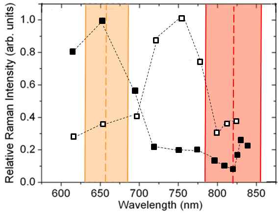
- Fig. 1: Evolution of the relative SERS intensity for nanocylinder shape excited with 632,8 nm line (black squares) and 785 nm line (white squares). The range [λ0-λRaman] for each excitation wavelength is colored.
[1] J. Grand et al. Synth. Met., 139, 621 (2003).
[2] J. Grand et al, Phys. Rev. B, 72, 033407 (2005)
[3] L. Billot et al., Chem. Phys. Lett., 422, 303 (2006)
[4] N. Guillot et al, Applied Physics Letters, 97, 023113 (2010)
University Paris XIII, CSPBAT Laboratory FRE 3043, 73 rue Marcel Cachin, Bobigny, France
H. Shen, and T. Toury
Université de Technologie de Troyes, Laboratoire de Nanotechnologies et d'Instrumentation Optique, Institut Charles Delaunay, CNRS-FRE 2848, 12 rue Marie Curie, Troyes, France
O. Peron and E. Rinnert
IFREMER, Service Interface et Capteurs, Département Recherche et Développements Technologiques, BP70, Plouzané, France
This study describes the effect of the surrounding medium on the SERS efficiency using nanolithographied substrates designed through electron beam lithography and lift off techniques. [1-3]
The investigation is done with liquid media gradually approaching the marine media compositions. Different compounds from probe molecule like trans-1,2-bis(4-pyridyl) ethylene (BPE) to Polycyclic Aromatic Hydrocarbons ( (PAHs) such as naphthalene are investigated. Their SERS intensities obtained from characteristic vibration bands are optimized as function of the surrounding medium in which they are immersed. The best SERS intensities are obtained by the optimization of nanolithographied substrates by choosing the appropriate gold nanoparticles size (Fig. 1). We will notably show that the optimum nanocylinder size is shifted to lower diameter by increasing the dielectric constant of the liquid medium. This rule will be discussed in term of Localized Surface Plasmon Resonance since its position influences directly SERS intensity.
This study is done for two excitation wavelengths, 632.8 nm and 785 nm, permitting the comparison of SERS substrates behaviour in both cases. The aim of this work is to collect some key information in order to product future active SERS sensors suitable for in situ environmental analysis.
The authors want to acknowledge the French National Agency (ANR) 07-P2IC-002-Discomar project, the Nanoantenna collaborative European project (HEALTH-F5-2009-241818) and the Conseil Régional de Champagne Ardenne for financial support.
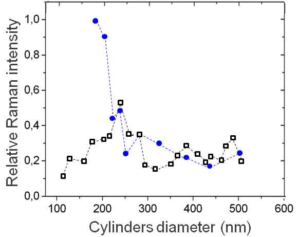
- Fig. 1: Evolution of the relative SERS intensity for nanocylinder shape excited with 785 nm line in air (white squares) and in water (full circles).
Institute of Photonics and Electronics, Chaberska 57, Prague, Czech Republic
homola@ufe.cz
Optical biosensors based on spectroscopy of surface plasmons propagating along metal-dielectric waveguides present the most advanced and mature optical label-free biosensor technology. Over the past fifteen years, SPR biosensors have become a central tool for characterizing and quantifying biomolecular interactions and have been also increasingly applied to detection of chemical and biological species [1]. SPR biosensor technology has been also commercialized by several companies.
This paper reviews recent advances in the SPR method, instrumentation, and applications. Selected advances in the SPR method to be discussed include, for instance, multiple-plasmon spectroscopy for bioanalysis of complex samples [2], polarization-contrast SPR imaging for high-performance parallelized SPR biosensing [3], and a new approach to spectroscopy of surface plasmons allowing construction of compact low-cost SPR sensing devices [4]. Performance of SPR sensors based on spectroscopy of propagating surface plasmons is discussed and compared with that of sensors utilizing localized surface plasmons [5]. Special microfluidic approaches for SPR biosensors are also presented. Approaches to immobilization of biorecognition elements (e.g. antibodies, nucleic acids) are discussed, including microfluidics-assisted and DNA-assisted approaches. Examples of bioanalytical applications illustrating bioanalytical performance and potential of SPR biosensors are presented. These include, in particular, detection of analytes related to medical diagnostics (micro ribonucleic acids, hormones, antibodies), environmental monitoring (endocrine disrupting compounds), food safety and security (drug residues, bacterial pathogens and toxins) [6].
[1] J. Homola, Surface Plasmon Resonance Based Sensors, Springer, Berlin, 2006.
[2] J. Dostalek, P. Adam, P. Kvasnicka, O. Telezhnikova, and J. Homola, Optics Letters 32, 2903 (2007).
[3] M. Piliarik, L. Parova, and J. Homola, Biosensors & Bioelectronics 24, 1399 (2009).
[4] M. Piliarik, M. Vala, I. Tichy, and J. Homola, Biosensors & Bioelectronics 24, 3430 (2009).
[5] P. Kvasnicka and J. Homola, Biointerphases, 3, FD4-FD11 (2008).
[6] J. Homola, Chemical Reviews 108, 462 (2008).
1Institute of physics and Center for Interdisciplinary Nanostructure Science and Technology - CINSaT, University of Kassel, Germany
2Institute of Optics and Atomic Physics, Technical University of Berlin, Germany
Surface enhanced Raman spectroscopy (SERS) is a powerful technique in the field of molecular spectroscopy. SERS exploits the superior optical properties of metal nanoparticles and allows single molecule detection. However, stable and reproducible SERS substrates, which are necessary for routine applications, are rarely available. Furthermore, to achieve an optimum enhancement for given molecule / excitation wavelength combinations, tailoring of the optical properties is desired. To meet these requirements, tailor-made SERS substrates were prepared, using silver or gold nanoparticles on sapphire supports. The tailoring is based on control of the mean shape and size of the nanoparticles, which permits a precise tuning of their localized surface plasmon polariton resonance in the vicinity of the excitation wavelength, used for the SERS measurements. In addition, the technique has been applied to study the influence of the wavelength on the SERS spectrum of pyrene.
In this contribution I present SERS measurements of pyrene as a function of the excitation wavelengths. In a first set of experiments I demonstrate, that neutral atom deposition, followed by subsequent diffusion and nucleation, i.e., Volmer Weber growth, is an suitable method to generate optimised SERS substrates. These substrates are highly reproducible and exhibit SERS enhancement factors in the order of 106, which are sufficient high for routine applications. In the second part of my talk, I will show the influence of the wavelength on the SERS spectra of pyrene. For this purpose, three different excitation wavelengths have been applied: λ = 514.5 nm, λ = 655 nm, and λ = 785 nm (cf. figure 1). Although in all experiments pyrene is clearly identified, only for the excitation wavelength of λ = 785 nm, a nearly background free Raman signal with an excellent signal-to-noise ratio is achieved.
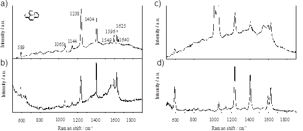
- Figure 1: Raman spectrum of bulk pyrene a). SERS spectra of pyrene obtained with an excitation wavelength of λ = 514.5 nm b), λ = 655 nm c), and λ = 785 nm d).
AG Biophotonik, FB Physik, Philipps-Universität Marburg, Marburg, Germany
Contact: dominik.huehn@physik.uni-marburg.de
Gold nanoparticle (AuNP) assisted remote-controlled release of immobilized molecules out of layer by layer (LBL) deposited polyelectrolyte interfaces [1] might be a promising application for drug delivery approaches. For that the recently thoroughly investigated characteristics of the light to heat conversion of gold nanoparticles/clusters upon their surface plasmon resonance (SPR) and the heat transfer to the surrounding medium might play the essential role [2,3].
This work concentrates on the characterization of polyelectrolyte multilayers assembled via the LBL technique, the embedding of functional molecules, and finally the excitation of these films within a near infrared (NIR) laser-modified microscope setup under various conditions. It is shown that both the extent of film destruction and the properties of arising vapor bubbles depend strongly on the medium the excitation takes place in.
[1] G. Decher, Science 277, 1232 (1997).
[2] A. O. Govorov, H. H. Richardson, Nano Today 2, 30 (Feb, 2007).
[3] C. Hrelescu et al., Journal Of Physical Chemistry C 114, 7401 (Apr 29).
Institute of Photonic Technology (IPHT) Jena, Nano Biophotonics Department
Sensor devices for bio analytic applications require high sensitivity and high specificity as well as high sensor density combined with an easy readout technology. Sensitivity and specificity depend on the specific biosensor whereas the other two requirements depend on the sensor device. Nanoholes in a thin metal film containing noble nanoparticles provide these features. Our present work focuses on basic investigations to proof the concept of this combined nanostructure. This includes correlated measurements on the combined structure with SEM, AFM, optical imaging and spectroscopy, in order to elucidate and characterize the parameters the signal is depending on. Such parameters are the depth of the hole, its diameter, the size of the particles in the hole, their number in the holes and, finally, the material of the particles. Some of these parameters can vary statistically over a wide range in a single probe. The result is a large variation of the individual signal of one nanohole. However, with statistical methods it is possible to extract single features which correlate with the parameters. The analysis allows for predictions of e.g. the number of particles in the holes or the size of the particles. Furthermore, the detection of changes in the environment is possible. This is shown for the binding of biomolecules on the surface. These measurements show a strong detectable optical signal, which is visible both in the optical images and the measured spectra. The signal is directly accessible from the optical images and, thus, provides an easy optical readout. The measurements can be automated to reach a high throughput with a suitable detector device. Based on this knowledge, a sensor device for bio analytic applications can be developed.
1 Institute of Photonic Technology, Jena, Germany
2 Dublin City University, Dublin, Ireland
This work reports about a novel label-free optical method for the detection of molecules based on a modification of localised surface plasmon resonance (LSPR) sensing by placing the metal nanoparticle close to the metal substrate. The optical properties of the nanoparticle are then significantly changed; plasmon resonance modes ('gap modes') are created and the scattering profile from the nanoparticles is altered. These effects strongly depend on metal nanoparticle metal substrate separation and gives better tenability to the system.
For the novel detection scheme gold substrate was used, which was covered with gold nanoparticles. Spectra of several nanoparticles were collected and topologies of the nanoparticles were recorded by atomic force measurement. The gold nanoparticles were then sequentially covered by ultra-thin (few nm thick) polymer layers applied by layer-by-layer deposition technique. After each layer, the nanoparticles' spectral properties were then recorded and the spectral shift of the LSPR was observed. Furthermore, the samples were incorporated into microfluidic chips and the absorption of the layers was detected in situ. The large sensitivity and tunability of this system indicate its applicability into bio-sensing platforms.
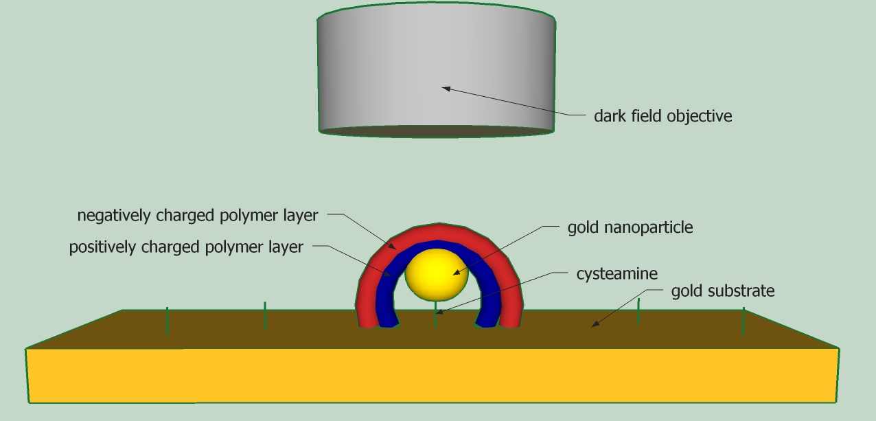
- Figure 1: Schematic of the dark field microscopy of a nanoparticle on a gold substrate covered with polymer layers
1Humboldt-Universität zu Berlin, Brook-Taylor-Str. 2, 12489 Berlin
2 Federal Institute for Materials Research and Testing (BAM), Richard-Willstätter-Str. 11, 12489 Berlin
Planar, nanostructured SERS substrates have become a basic tool for applications of SERS to analytical problems (1), as such a substrate permits reproducibility of SERS signals from an analyte. Here, we report on the construction of dedicated, planar SERS sensors based on immobilization of nanoparticles on glass substrates via polymers. In particular, the electromagnetic contribution to the SERS enhancement of individual gold nanospheres that are used as basic building blocks for the nanostructured surface was determined experimentally (2). So far, experimental proof of the theoretical predictions (3) of such enhancement factors was still missing. Immobilization and functionalization of the nanoparticles is a crucial step in the construction of the sensor. For the characterization of the properties of the nanoparticles in solution and on the substrate, direct (electron microscopy, scanning force microscopy) and indirect methods (UV/Vis absorption) have been used. Furthermore the stability and reproducibility of the substrate and the influence of different parameters (e.g. concentration, functional group, properties of the nanoparticles in solution) during the assembly were examined. A major hallmark of our novel sensors is the possibility to combine nanostructures of different materials.
Using novel planar composite sensors, we show how SERS can be used for the determination of catalytic activity. The fast determination of catalytic activity is important in the development of new catalysts. For hydrogenation reactions the conversion of 4 nitrophenol to 4 aminophenol by sodium borohydride is used as model reaction. Usually, the reaction is monitored by UV/Vis spectroscopy which is very time consuming. In contrast, the application of SERS is very fast and molecule-specific.
The use of an internal standard allows a relative quantification and the determination of reaction rates.
So far, this sensor was used with different catalysts such as platinum nanoparticles, copper acetylacetonat and Raney-nickel. Future applications include studies in the area of biocatalysis.
(1) Wunder, S.; Polzer, F.; Lu, Y.; Mei, Y.; Ballauff, M. J. Phys. Chem. C 2010, 114, 8814-8820.
(2) Joseph et al Journal of Raman Spectroscopy (in press)
(3) Zeman, E.J.; Schatz, G.C. J. Phys. Chem. 1987, 91, 634-643
1Department of Biochemistry, George S. Wise Faculty of Life Sciences, Tel Aviv University, Ramat Aviv, 69978 Israel,
2Nanotechnology Center, Tel Aviv University, Ramat Aviv, 69978 Israel
We have recently reported that incubation of 20 nm gold nanoparticles (Au-NPs) with G-quadruplex molecules results in the formation of stable G4-DNA-coated nanoparticles and DNA-NP conjugates [1]. Incubation of Au-NPs with parallel stranded G-quadruplexes, comprising phosphorothioate residues on both sides of the DNA, also yields stable nanoparticles. The particles are covered with phosphorothioate groups that can anchor them to metal surfaces and other metal particles. Incubation of these "sticky" particles with 10-fold excess of 20 nm bare (citrate-coated) Au-NPs leads to the formation of flower-shaped structures (NP-flowers) comprising a central non-coated particle and several G-quadruplex coated ones at the periphery (see Fig. 1A). Absorption spectra of the structures are presented in Fig.1B. The absorption band of AuNPs is shifted towards long wavelengths compared to individual particles not connected to each other (compare curve 1 and 4 in Fig. 1B). The spectral shift depends on the length of the DNA molecules connecting core particle with peripheral ones in the NP-flower. A pronounced shift is observed for the 5 tetrad G-quadruplex (curve 4 in Fig. 1B), the effect is less pronounced for the 10 tetrad one (Fig. 1B, curve 3) and can hardly be seen for the 20 tetrad DNA (Fig.1B, curve 2). This result shows the strong dependence of plasmon coupling strength on the distance between metal particles in the 1.5 nm (the length of the 5 tetrad quadruplex molecule) - 6 nm range. We have also shown that the absorption spectrum of Au-NP dimers, in which the two particle are connected by 10 tetrad G-quadruplex linkers is similar to that of single particles. We therefore conclude that the observed plasmon resonance shifts (see Fig 1B) are due to cooperative interactions and depend on the amount of AuNPs in the structure.
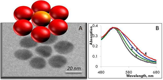
- Figure 1. TEM and absorption spectroscopy of NP-flowers. A - TEM image (bottom) and schematic presentation (top) of the NP-flower. In the flower a central 20 nm AuNP is surrounded by six AuNPs of the same size. B – Absorption spectra of 20 nm AuNP (curve 1) and NP-flowers, in which particles are connected to each other by 20 (curve 2), 10 (curve 3) and 5 (curve 4) tetrad G-quadruplex molecules.
1. Lubitz I., Kotlyar A. Self-assembled G4-DNA-silver nanoparticle structures. (2011) Bioconjug. Chem., 22, 482-487.
University of Maryland School of Medicine
Center for Fluorescence Spectroscopy
Department of Biochemistry and Molecular Biology
725 W Lombard St, Baltimore, Md 21201
Fluorescence spectroscopy is widely used in chemical and biological research. Until recently essentially all fluorescence experiments were performed in the far-field regime. By far-field we mean at least several wavelengths from the fluorophore. In recent years there has been a growing interest in the interactions of fluorophores with metallic surfaces or particles. Near-field interactions are those occurring within a wavelength distance of an excited fluorophore.
The spectral properties of fluorophores can dramatically be altered by near-field interactions with the electron clouds present in metals. These interactions modify the emission in ways not seen in classical fluorescence experiments. Fluorophores in the excited state can create plasmons that radiate into the far-field and that fluorophores in the ground state can interact with and be excited by surface plasmons. These reciprocal interactions suggest that the novel optical absorption and scattering properties of metallic nanostructures can be used to control the decay rates, location, and direction of fluorophore emission. We refer to these phenomena as plasmon-controlled fluorescence (PCF). We will present a review of the recent work on metal-fluorophore interactions and suggest how these effects could result in new classes of experimental procedures, novel probes, bioassays and devices. While there are many complexities to fluorophore-metal interactions there are also many opportunities. In addition to control of the chemical structure of the fluorophore we can now control, to some extent, the processes of excitation and emission with nearby metallic nanostructures resulting in new approaches to high-throughput biology, diagnostics and molecular imaging.
1Universite Paris XIII, Laboratoire CSPBAT (FRE 3043), UFR SMBH, equipe LBPS, 93017 Bobigny, France
2Universite de technologie de Troyes, Laboratoire de Nanotechnologie et d'instrumentation Optique, Institut Charles Delaunay, FRE 2848, 12 rue Marie Curie, 10010 Troyes, France
email: marc.lamydelachapelle@univ-paris13.fr
Our SERS sensor is based on gold nanocylinder arrays obtained by Electron Beam lithography. The preliminary results concern the first study of SERS efficiency of nanocylinder arrays at 632 nm in the case of proteins, BSA and RNAse. The maximum SERS intensity obtained for a specific nanocylinder diameter.
Surface-enhanced Raman scattering (SERS) technique has proved to be a very effective analytical tool of molecules due to its high sensitivity. It has been widely used for ultrasensitive chemical analysis down to single molecule detection [1], and it expands its field of applications from chemical-biochemical analysis to nanostructure characterization and biomedical applications [2,3]. It is very useful in detecting conformational changes and structural differences regarding preferred orientations of molecules with respect to a metal surface [4, 5].
The purpose of the present study is to achieve a SERS sensor with extreme sensitivity and very high molecular specificity by combining the enhancement of vibrational signal, surface functionalization, respectively, and detection of vibrational Raman spectra signals in order to provide at very low concentration the spectral signature of the molecule to detect. This should allow a significant improvement in sensor performance, including in terms of reliability and speed detection. Our SERS sensor is based on gold nanostructures obtained by Electron Beam lithography. This technique makes possible the manufacture of highly reproducible patterns containing particles with different geometries, shapes and size. These metal nanostructures seem to be suitable substrates for the study of SERS mechanism. It is known that the size and shape of these arrays of these particles should be optimised so as to give rise to the SERS spectrum of a molecule adsorbed at very low concentration. And in this case the plasmon band is suitably located with respect to the laser excitation wavelength [6].
In the present study the arrays of particles are nanocylinder with different diameters.
Our preliminary results concern the first study of SERS efficiency of nanocylinder arrays at 632 nm in the case of proteins, BSA (1mM) and RNAse (1mM). In the case of the BSA the protein adsorption on arrays structure does not induce conformational changes while in the case of the RNAse structural changes are evident (figure 1). The maximum SERS intensity is obtained for a specific nanocylinder diameter (100nm). These results give evidence that the nanocylinder size should be optimised in order to obtain highly enhancement factor.
For the two proteins the enhancement factors (measured for the bands located at 1616 cm-1 for RNAse and 1660 cm-1 for BSA) were estimated to 106-107. These values are coherent with those obtained by Felidj and co-workers for the cylinder gold structure [6].
Acknoledgments:
This work is supported by FP7-HEALTH-F5-2009-241818 - NANOANTENNA European project.
[1] K. Kneipp, Y. Wang, H. Kneipp, L. T. Perelman, I. Itzkan, R.R. Dasari and M. S. Feld, Phys. Rev. Lett. 1997, 78, p1667 - 1670.
[2] R. Aroca, Surface-Enhanced Vibrational Spectroscopy; John Wiley & Sons Ltd.: Chichester, UK, 2006, p141-176.
[3] K. Kneipp., M. Moskovits, H. Kneipp, Eds. Surface-Enhanced Raman Scattering: Physics and Applications; Springer: Berlin, 2006
[4] K. E. Shafer-Peltier, C. L. Haynes, M. R. Glucksberg, R. P. Van Duyne, J. Am. Chem. Soc. 2003, 125, p588-593
[5] X. L. Li, W. Q. Xu, J. H. Zhang, H. Y. Jia, B. Yang, B. Zhao, B. F. Li, Y. Ozaki, Langmuir , 2004, 20, p1298-1304
[6] N. Felidj, J. Aubard, and G. Levi, Physical Review B, 2002, 65, 075419
1Institute of Photonic Technology Jena, 07745 Jena, Germany
2Max Planck Institute for the Science of Light, 91058 Erlangen, Germany
3Department of Solid State Electronics and CeNIDE, University of Duisburg-Essen, 47057 Duisburg, Germany
Electrical and optical properties of semiconducting nanowires (NWs) are dependent on their diameters. Hence an exact knowledge about the diameters of NWs during device processing is fundamental to control their unique properties. Here we present a precise optical method to easily determine the diameters of individual NWs with a relative error less than 10%. This method has the advantages that no vacuum technique is needed, only relatively cheap hardware is used and that it has the potential to be scaled up. The underlying physical principle are strong Mie resonances which dominate the optical scattering spectra of most semiconducting NWs. Our findings show that even slight tapering of the NWs can be detected in an optical microscope. Also step like diameter changes of a few nanometers, as they occur when the growth conditions are changed, can be clearly identified. In addition a profound analyses of the elastic scattering properties of individual semiconducting NWs is presented.
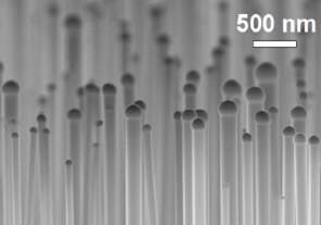
- VLS grown GaAS-Nanowire
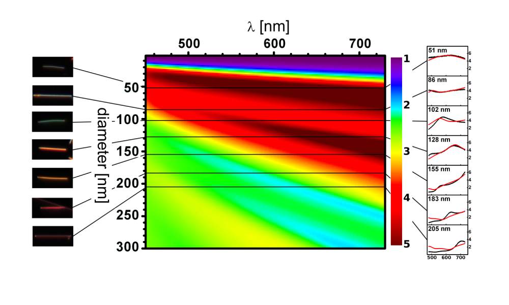
- Scattering efficiency of GaAs-Nanowire
Science Institute, University of Iceland, Dunhagi 3, IS107 Reykjavik, Iceland
We are currently investigating methods of integrating plasmonic structures with high-index-contrast polymer waveguides, in order to combine plasmonic sensing with efficient on-chip transmission and manipulation of optical signals. In particular, we have studied long-range surface plasmon polariton (LRSPP) waveguides with a symmetric cladding environment. The symmetric environment is provided on one side by an aqueous sample and a fluorinated optical polymer with a similar refractive index (n=1.34) on the other [1]. We have previously realized evanescent-wave fluorescence excitation in fixed cells [2] and various highly integrated polymer optical devices [3] using this cladding material. Strong coupling between a plasmonic and a dielectric waveguide can serve to transmit excitation energy to the sensing region and/or to couple SPP-quenched fluorescence to propagating modes in the dielectric waveguide.
In addition, we will discuss recent developments in using fluorescent polymers for realizing optical gain in plasmonic waveguides [4]. The combination of plasmonic structures and semiconducting polymers, such as derivatives of poly(phenylene vinylene) or poly(fluorine) co-polymers, represents an interesting avenue of research in molecular plasmonics since these polymers can provide substantial optical gain in the visible range, close to the surface plasmon resonance of typical plasmonic metals. This is especially true for LRSPP waveguide configurations where propagation losses can be reduced below the value of the achievable optical gain, providing net amplification of propagating plasmons in the visible range.
[1] B. Agnarsson, J. Halldorsson, N. Arnfinnsdottir, S. Ingthorsson, T. Gudjonsson, K. Leosson, Microelectronic Engineering 87, 56 (2010)
[2] B. Agnarsson, S. Ingthorsson, T. Gudjonsson, K. Leosson, Opt. Express 17, 5075 (2009)
[3] J. Halldorsson, N.B. Arnfinnsdottir, A.B. Jonsdottir, B. Agnarsson, K. Leosson, Opt. Express 18, 16217 (2010)
[4] M.C. Gather, K. Meerholz, N. Danz, K. Leosson, Nature Photonics 4, 457-461 (2010)
1 Karlsruhe Institute of Technology (KIT), Institute for Microstructure Technology, D-76128 Karlsruhe, Germany
2 Institute of Photonic Technology (IPHT), Nanophotonic Department, D-07702 Jena, Germany
email: timo.mappes@kit.edu
This talk will provide an insight into the microscope equipment used in the very beginning of ultramicroscopy of colloids in both, liquid and solid media.
Ultramicroscope for colloids after H. Siedentopf and R. Zsigmondy
Colloidal fluids were examined with the slit ultra microscope introduced by R. Zsigmondy. W. Biltz suggested an improved cuvette that was connected to a bowl for the colloids to be examined. The entire setup was then mounted directly with a holder to the water immersion objective D* (40x, N.A. 0.75) by Carl Zeiss Jena (Fig. 1).
Ultramicroscopy of living bacteria
Living bacteria could be examined with the help of a cardioid condenser (available in glass and quartz) and two dedicated apochromatic objectives. A condenser and one of these objectives are illustrated in Fig. 2.
Immersion Ultramicroscope after R. Zsigmondy, improved by A. Ehringhaus
First introduced in 1913 by R. Zsigmondy and patented with using two water immersion objectives (6.2 mm, N.A. 1.05) the Immersion Ultramicroscope was improved by A. Ehringhaus in 1919 and fabricated by R. Winkel in Göttingen. The 1930ies version of this particular stand is shown in Fig. 3.
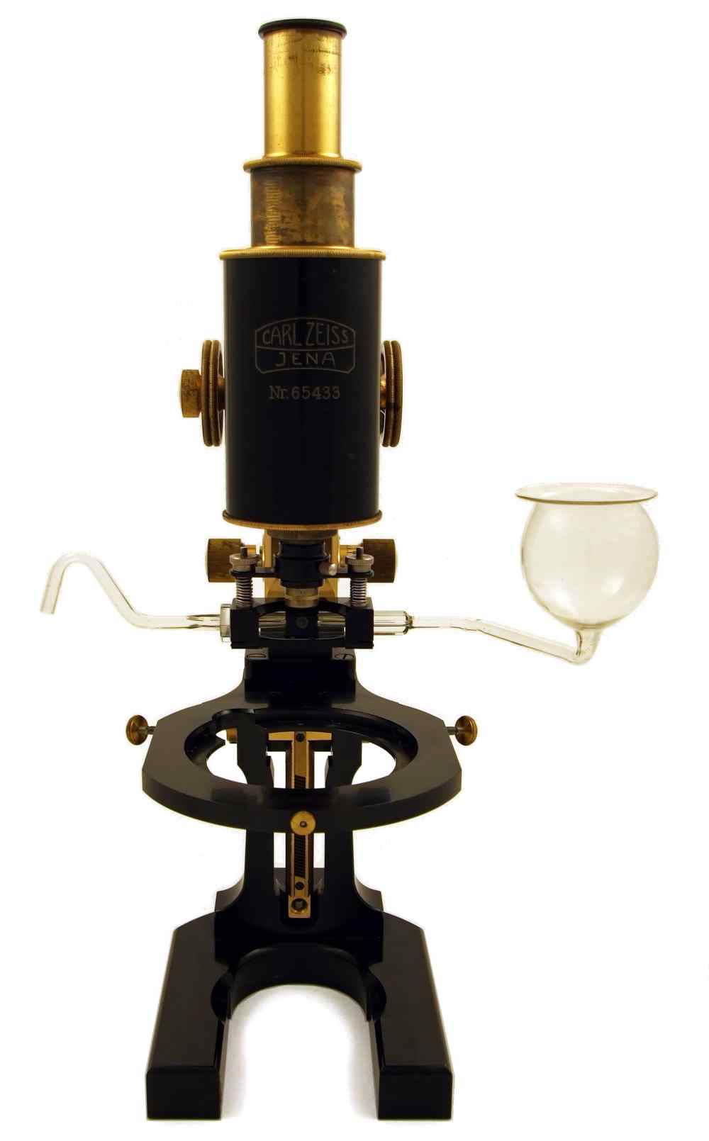
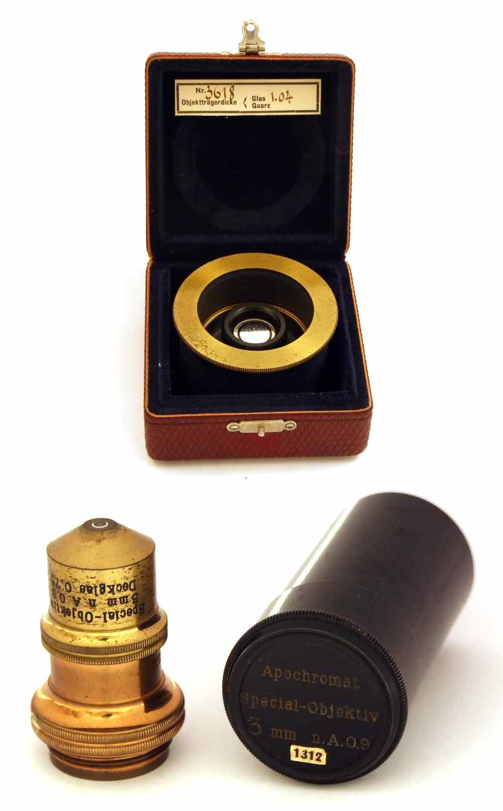
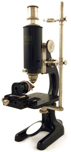
Fig.1(left): Ultramicroscope outfit by Carl Zeiss Jena: Large research microscope stand of 1914 with cuvette after W. Biltz in fitting mounted with water immersion objective D* (40x, N.A. 0.75). Fig. 2.(middle): Ultramicroscopy accessories by Carl Zeiss Jena pre 1920: Cardioid condenser and Apochromat (3 mm, N.A. 0.9) for ultramicroscopy of living bacteria. Fig. 3.(right): Patented Immersion Ultramicroscope by Winkel-Zeiss, No. 32607 of 1930.
Philipps Universität Marburg, 35037 Marburg, Germany
In recent years, many delivery systems for biological or medical applications have been developed. The use of nano- or microcontainers such as liposomes or polyelectrolyte microcapsules has reached great interest in this field. Nanoparticle functionalized microcapsules fabricated via Layer-by-Layer (LbL) adsorption of polyelectrolyte materials and charged gold nanoparticles1,2 are one promising approach to perform remote-controlled release of material inside living cells or tissues3. Our research benefits from plasmon-assisted photothermal processes, in which light absorption and heat dissipation are competing mechanisms that rule the actual temperature in metallic nanostructures embedded within the microconainers.
Herein, a near-IR laser diode serves as light source for energy transfer. The laser emission wavelength of 830nm is crucial, as its power is mainly passing the biological matter without any interaction. Though, the surface plasmon resonance peak of (spherical) gold-nanoparticles is around 520nm. Therefore clusters of gold or rodshaped particles have been used for this approach4.
Release of different macromolecules conjugated with organic fluorophores from so-modified capsules has been proven via fluorescent microscopy and electron microscopy imaging.
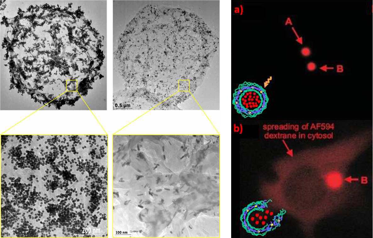
- Fig. 1 (left): Empty multilayer microcapsules fabricated by subsequential absorption of polyelectrolytes onto spherical templates functionalized with clusters of gold nanoparticles (left) and gold nanorods (right). Fig. 2 (right): (a) Cell containing microcapsules filled with AlexaFlour594 before treatment with IR-laser beam. (b) After heat formation via laser treatment, fluorophore is released from the capsule and distributed over the cytosol of the cell.
1 Mercato, L. L. d.; Rivera-Gil, P.; Abbasi, A. Z.; Ochs, M.; Ganas, C.; Zins, I.; Sönnichsen, C.; Parak, W. J. Nanoscale 2010, 2, , 458-467
2 Munoz-Javier, A.; Pino, P. d.; Bedard, M.; Skirtach, A. G.; Ho, D.; Sukhorukov, G.; Plank, C.; Parak, W. J. Langmuir 2009, 24, 12517-12520
3 Skirtach, A. G.; Javier, A. M.; Kreft, O.; Köhler, K.; Alberola, A. P.; Möhwald, H.; Parak, W. J.; Sukhorukov, G. B. Angew. Chem. Int. Ed. 2006, 45, 4612-4617
4 Skirtach, A. G.; Karageorgiev, P.; De_Geest, B. G.; Pazos-Perez, N.; Braun, D.; Sukhorukov, G. B. Advanced Materials 2008, 20, 506-510
Philipps Universität Marburg, Fachbereich Physik, Renthof 7, 35037 Marburg, Germany
Email: christian.pfeiffer@staff.uni-marburg.de
The synthesis and the study of the behavior of sliver nanoparticles (Ag NPs) for example in cells is very important because the number of commercial product containing Ag increase every day. Ag NPs were synthesized in organic solvent (ethanol) carrying alkanethiolate ligands at their surface [1]. To make these particles water dispersible they must be coated with an amphiphilic polymer [2]. The polymer used was poly(isobutylene-alt-maleic anhydride), modified with dodecylamine. The modification of the particles with dyes or organic sensors, permits us to monitor the behavior of these particles in the cells or use them as nanosensors.
The concentration of Ag NPs solutions were determined by UV/vis spectroscopy and Lambert-Beers law. Therefore the extinction coefficient of the Ag NPs was calculated by the method of Graham et al. [3].
[1] Mari A., et al.; High yield synthesis of pure alkanethiolate-capped silver nanoparticles; Langmuir, 2010, 26(19), 15561-15566.
[2] Cheng-An J. L., et al.; Design of an amphiphilic polymer for nanoparticle coating and functionalzation; Small, 2008, 4(3), 334-341.
[3] Graham A., et al.; Extinction coefficient analysis of small alkanethiolate-stabilised gold nanoparticles; Chemical Physics Letters, 2008, 460, 230 - 236.
1Belarusian National Technical University, Minsk, pr. Independence 65, 220013, Belarus.
pustovalovv@mail.ru
2Stepanov Institute of Physics, National Academy of Sciences of Belarus, pr. Independence 68, 220072 Minsk, Belarus
Applications of plasmonic nanoparticles (NPs) as photothermal (PT) agents for different applications in laser nanotechnology and nanomedicine are fast growing areas of research. Many potential benefits include possibility for imaging and treatment of materials containing of NPs, applications of NPs in catalysis, medicine, chemistry, etc. Efficiency of applications of metallic nanoparticles for laser and optical nanotechnology depend on optical and thermo-physical properties of NPs and characteristics of radiation. Nevertheless, despite successful results, there is a lack of focused analysis of requirements to NPs properties for optimization of PT applications, especially with pulsed lasers. Here we present a platform for comparative analysis of NP properties (e.g., optical, thermal, structural, and others) allowing to select their parameters for concrete technology. Parameter ΔT0/E0 [1, 2]
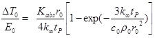
can be used for determination of thermo-optical properties of NPs, where ΔT0=Tmax- T∞, T∞ - initial temperature, maximal temperature of NP Tmax = T0(tP), ρ0 , c0 - density and heat capacity of NP material respectively, r0 - radius of a spherical NP, Kabs (r0, λ,) is the efficiency absorption factor of radiation with wavelength λ by a spherical particle of radius r0 [3, 4], k∞ - coefficient of thermal conductivity of a surrounding medium, tP - laser pulse duration. Parameter (1) may be viewed as NP heating efficacy depending on r0 , Kabs (λ), ρ0 , c0 , tP and k∞ under action of radiation energy density E0. This parameter determines the increase of NP temperature under action of laser radiation with energy density value is equal 1 J/cm2.
The thermo-optical properties of several metallic (aurum, silver, platinum, cobalt, zink, nickel, titanium, etc.) NPs are theoretically investigated and compared which provide significant increased conversion of laser pulse energy in PT phenomena. Some of these NPs make it possible to use them with maximal efficiency in laser and optical nanotechnology and nanomedicine.
Authors would like to thank Belarusian foundation of fundamental research for support.
[1] Pustovalov, V.K.; Smetannikov, A.S.; Zharov, V.P. Photothermal and accompanied phenomena of selective nanophotothermolysis with gold nanoparticles and laser pulses. Las. Phys. Lett. 2008, 5, 775-792.
[2] Pustovalov V, Astafyeva L, Jean B Computer modeling of the optical properties and heating of spherical gold and silica-gold nanoparticles for laser combined imaging and photothermal treatment. Nanotechnology, 2009, 20, 225105.
[3] Bohren, C.F., Huffman, D.R. Absorption and Scattering of Light by Small Particles; Wiley: New York, 1983.
[4] Pustovalov, V.K.; Babenko, V.A. Optical properties of gold nanoparticles at laser radiation wavelengths for laser applications in nanotechnology and medicine. Las. Phys. Lett. 2004, 1, 516-520.
COSINGO, Imagine Optic Spain SL, Castelldefels (Barcelona), Spain,
email: frohde@cosingo.com
Summary
We present the prototype of a biosensor that exploits surface plasmon resonances of gold nanostructures integrated in a
microfluidic environment to track HSP70 proteins in the peripheral blood.
Introduction
Surface plasmon resonances (SPR) have become a standard tool for performing concentration analysis of biological samples,
determining affinity constants of biomolecules or measuring chemical reaction kinetics. Commercial SPR systems are widely
available and can be found in many modern bio-chemistry labs. Recent experiments suggest that using nanostructures exhibiting
localized surface plasmon resonances (LSPR) could be the basis for biosensors of comparable or even better performance [1].
SPEDOC (Surface Plasmon Early Detection of Circulating Heat Shock Proteins) is a research initiative supported by the European
Comission's 7th Framework programme that aims at combining the latest advances of nano-optics, optical manipulation and microfluidics
with recent discoveries about Heat shock Proteins (HSP) to develop the precursor of future individualized cancer diagnosis and
treatment follow-up devices.
COSINGO is a company that unifies great expertise in optical engineering, in applied research with optical technologies and
long experience building optical sensors. For SPEDOC, COSINGO teams up with four research institutions from France, Switzerland
and Spain; ICFO, UB, INSERM and EPFL.
Discussion
The platform developed during this project will exploit the surface plasmon resonances supported by gold nanostructures
integrated in a microfluidic environment to track HSP70 proteins in the peripheral blood. This innovative sensor should
permit providing treatment to cancer patients at an earlier stage and at lower doses with the consequent decrease of secondary effects.
We will present the project objectives and show first results achieved with the first generation prototype of an optical detection
platform (ODP) developed by COSINGO. This ODP is designed for the detection of proteins in solution and uses a PDMS microfluidic
chip of the type shown in figure 1. The setup will be presented and results of the first experiments measuring the plasmon resonance
shift of nanodimers (illustrated in figure 2) when exposed to solutions of different refractive indices will be presented.
The measurements are performed by data acquisition software using the centroid tracking method [2]. This allows for live monitoring
of binding dynamics and greatly improves the sensitivity of the associated sensor.
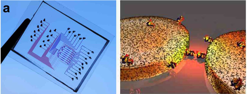
- Fig 1 (left). First generation micro-fluidic chip. Fig 2( right). Illustration of functionalized nanodimers.
[1] A.B. Dahlin, S. Chen, M.P. Jonsson, L. Gunnarsson, M. Käll, F. Höök, Analytical chemistry, 81 (2009) 6572-6580.
[2] A.B. Dahlin, J.O. Tegenfeldt, F. Höök, Analytical chemistry, 78 (2006) 4416-4423.
1Institut Fresnel, CNRS, Aix Marseille Université, École Centrale Marseille,
Domaine Universitaire de Saint Jérôme, 13397 Marseille
2Institut Langevin, CNRS, ESPCI,
10 rue Vauquelin, 75005 Paris
*corresponding author. Email adress : nicolas.bonod@fresnel.fr
When excited in the near field by a quantum source of light, optical antennas allow (i) to increase the decay rate of these
emitters, thus increasing the number of emitted photons per second, but also (ii) to redirect the emitted radiation in a narrow
angle, yielding a better collection efficiency. This latter property, which is of primary importance, will be the subject of this talk.
First, we will show that a metallic particle coupled to a dipolar emitter can strongly modify the radiation pattern. For example,
when the emitter is placed at 30nm of the center of a silver spherical particle, 85% of its radiation at 600nm is emitted in a
single half-space. But we will evidence a new phenomenon : when the distance is increased by 6nm, only 50% of the power is scattered
in the same half-space, as the radiation pattern is symmetric.
We will show that this symmetry results from a phase opposition between the emitter (fluorescent molecule, quantum dot) and the
induced dipole (metallic particle). Then, we will show that this phase difference depends on 3 terms : (1) the particle polarizability
phase, (2) the far-field propagation term, and (3) the near-field coupling of the dipoles. We will analyse the evolution of the two
latter terms in function of the emitter/particle distance and evidence the crucial role of the near-field term. We will finally
explain why a 6nm modification in the distance results in such a strong modification of the radiation pattern.
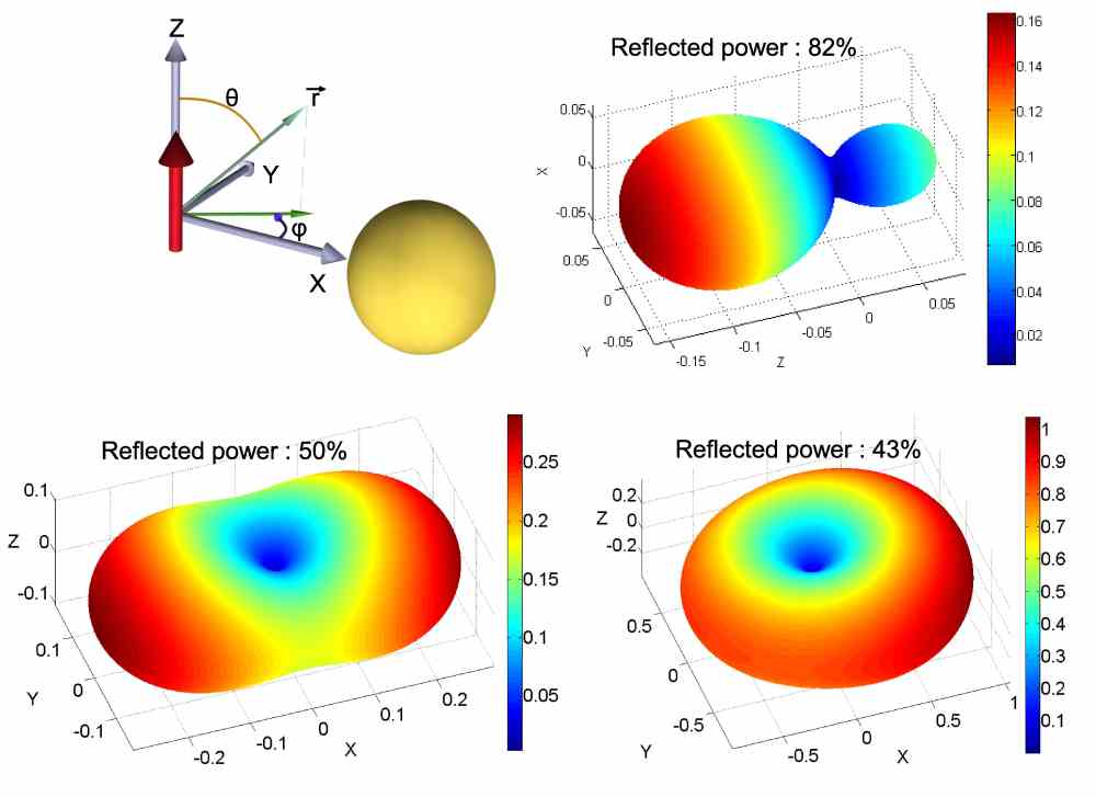
- Radiation pattern of a dipolar emitter placed at (top right) 30nm, (bottom left) 36nm, and (bottom right) 76nm of the center of a spherical particle 50nm in diameter. λ0=600nm, background medium n=1.5.
1. N. Bonod et al., "Ultracompact and unidirectional metallic antennas," Phys. Rev. B 82, 115429 (2010).
2. B. Rolly, et al. "Crucial role of the emitter-particle distance on the directivity of optical antenna," submitted.
Institute of Photonic Technology (IPHT) Jena, Germany
thomas.schneider@ipht-jena.de
Early understanding of the physics of nanoparticle plasmons dates back to Faraday [1] and Mie [2], and the extraordinary optical
properties has been the subject of extensive studies [3]. Noble metal nanoparticles exhibit a strong UV-visible extinction band
which is not present in the spectrum of bulk material [4]. This phenomenon is known as localized surface plasmon resonance (LSPR)
and the extinction band occurs when the incident photon frequency is resonant with the collective excitation of the conduction electrons.
It is well established that intrinsic parameters like size, geometrical shape, material and composition, but even more extrinsic
parameters such as charge distributions and dielectric properties of the nanoparticle's immediate environment strongly influence
the maximum peak wavelength of the LSPR spectrum [3, 4].
In this work we demonstrate the use of single gold nanoparticles as sensing platform for a label-free detection of hybridization
events of single-stranded capture oligonucleotides with complementary target sequence. Therefore, different reaction conditions
for like thiol-group pre-activation, the use of a poly A tail and pre-incubation with 6-mercaptohexanol for increasing the
hybridization efficiency were tested. Concentration dependence of the target DNA oligonucleotide could be shown by end point
and online measurements respectively. Also hybridization events of a DNA sandwich system containing a thiol-modified capture
oligonucleotide, a longer linker DNA molecule for binding of a third DNA strand was monitored. Sensitivity investigations of
different nanoparticles were done by replacing the gold nanospheres by other gold nanostructures like rings, split rings and discs.
The project was financially supported by the German Research Foundation DFG (FR 1348/12-1)
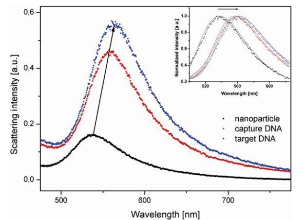
- Fig.1: DNA immobilization and sensing of the DNA hybridization event is shown as a red shift of the maximum wavelength. The black spectrum indicates the "naked" gold nanoparticle, the red spectrum shows the nanosphere after functionalization with the capture DNA and the blue spectrum displays the hybridization event. Spectra were fitted by an extreme fit function (grey lines).
[1] M. Faraday, Philosophical Transactions of the Royal Society of London, 147, 145-181 (1857)
[2] G. Mie, Annalen der Physik, 330 (3), 377-445 (1908)
[3] U. Kreibig, M. Vollmer, Optical Properties of Metal Clusters, Berlin, Springer (1995)
Physical Biology (FB 15, CEF-MC, FMLS), Goethe Universität, Frankfurt am Main, Germany
Ernst.Stelzer@PhysikalischeBiologie.de
Specimens scatter and absorb light, thus, the delivery of the probing light and the collection of the signal light become inefficient; many endogenous biochemical compounds also absorb light and suffer degradation of some sort (photo-toxicity), which induces malfunction of a specimen. In conventional and confocal fluorescence microscopy, whenever a single plane is observed, the entire specimen is illuminated (Verveer 2007). Recording stacks of images along the optical z-axis thus illuminates the entire specimen once for each plane. Hence, cells are illuminated 10-20 and fish embryos 100-300 times more often than they are observed (Keller 2008). This can be avoided by using light sheets, which are fed into the specimen from the side and overlap with the focal plane of a wide-field fluorescence microscope. In contrast to an epi-fluorescence arrangement, such an azimuthal fluorescence arrangement uses two independently operated lenses for illumination and detection (Stelzer 1994; Huisken 2004).
Optical sectioning and no photo-toxic damage or photo-bleaching outside a small volume close to the focal plane are intrinsic properties. Light sheet-based fluorescence microscopy (LSFM) takes advantage of modern camera technologies. LSFM can be operated with laser cutters (e.g. Colombelli 2009) and in fluorescence correlation spectroscopy (FCS, Wohland 2010). We have also successfully evaluated the application of structured illumination in a SPIM (Breuninger et al., 2007). Christoph Engelbrecht investigated the performance of our SPIMs (Engelbrecht & Stelzer, 2006) both theoretically and practically. We also designed and implemented a wide field frequency domain Fluorescence Lifetime Imaging (FLIM) setup, which is based on a SPIM. This arrangement provides an inherent optical sectioning capability and reduces photo bleaching compared to conventional wide field and confocal fluorescence microscopes. Most recently, we implemented incoherent structured illumination in our DSLM (Keller 2010). The intensity modulated light sheets can be generated with dynamic frequencies and allow us to estimate the effect of the specimen on the image formation process at various depths in objects of different age.
The single plane illumination microscope (SPIM) employs a cylindrical lens to generate a light sheet. A collimated laser beam is focused into the plane of the detection lens along one direction while the other direction remains collimated (Greger et al., 2007). While this approach is relatively simple and straightforward it suffers from the low quality of the cylindrical lens and the inefficiency of the illumination system (Breuninger et al., 2007). The major advantage of the digital scanned laser light sheet-based fluorescence microscope (DSLM; Keller et al., 2008) is that it relies entirely on cylindrically symmetric optics and hence provides a very good optical quality. In addition, the DSLM employs a minimal number of optical components and does not suffer from excessive wave front aberrations.
The development of LSFM at EMBL draws on many previous developments. In particular confocal theta fluorescence microscopy, which was originally developed together with Steffen Lindek played a very important role. About a dozen papers on theta microscopy describe its properties and that of LSFM (single & two-photon, annular/Bessel beams, (a)symmetric arrangements, …) theoretically as well as practically.
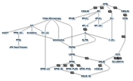
All references are found via: http://www.researcherid.com/rid/A-7648-2011
Institute of Photonic Technology, Albert-Einstein-Straße 9, 07745 Jena, Germany
In this report, presenting the research activities of our group in the field of metal nanoparticles, four different aspects
of use of metal nanoparticles (mNP) will be introduced. It will be shown that mNPs can be used for light concentration and
guiding as well as for generating a local light source with an efficient energy coupling and how these effects can be used
for sensing and nanofabrication applications.
Metal nanoparticles have the ability to strongly focus light around them at a specific wavelength. This effect is caused by collective
action of confined electrons in the mNP and is termed Localised Surface Plasmon Resonance (LSPR). The mNPs can be used as an effective
localized nanosensor for molecules absorbed to the NPs by monitoring the LSPR wavelength, because the LSPR wavelength depends on the
local polarizability around the mNP. In the first example we will present results of the detection of DNA hybridization by LSPR
measurements (Figure 1 A). LSPR wavelength of single mNPs was monitored by measuring spectrum of scattered light from the NPs in a
dark field microscopy set up. In the second example, the LSPR of mNP was modified by placing the mNP in the close proximity of a
metallic layer. This system focuses the light strongly in the gap between the mNP and the substrate and creates "gap modes". These
gap modes are very sensitive on the NP-substrate separation and we investigated their properties by single particle dark field
microscopy (Figure 1 B). Further, metallic nanoparticles were used to modify transmission properties of sub wavelength apertures.
We will present experimental data as well as theoretical calculations showing that a mNP placed in a hole in a chromium layer can
increase the light transmission through the hole at specific wavelengths (Figure 1 C). Last, we will reveal how mNP can be used as
localized energy source through femtosecond laser pulse illumination. Through non-linear processes the mNP acts as a source of higher
harmonics. This localized energy can be then efficiently coupled into the near-by laying DNA bundle and further propagates along the
DNA bundle. The experimental evidence of this coupling is demonstrated by destruction of the underlying substrate (Figure 1 D).
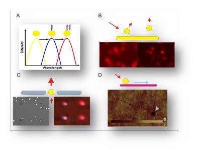
- Figure 1 A- Detection principle of DNA hybridization on a single NP by dark field microscopy. B – Dark field images of mNP with different distances from a metal substrate. The scattering profiles as well as spectra are strongly dependent on the separation. C – (left) TEM images of sub-wavelengths apertures with and without mNPs and (right) their corresponding transmission intensities. D – AFM image of a substrate destroyed by energy generated in the mNP and propagating along the DNA bundle after fs laser pulse illumination.
aGraduate School of Frontier Biosciences, Osaka University, Suita, Japan
bWPI Immunology Frontier Research Center, Osaka University, Suita, Japan
s-tanaka@ap.eng.osaka-u .ac.jp
Noble-metal nanoclusters (Au and Ag) consisting of several atoms have been gaining much attention as novel fluorescent markers due to their optical properties, similar to semiconductor quantum dots, such as size-dependent emission wavelength and discrete electronic state. Metal nanoclusters are supposedly superior to semiconductor quantum dots for their smaller size and less cytotoxicity. So far, several groups have reported biological application of noble-metal nanoclusters (Au and Ag) as fluorescent probes in cellular imaging. For instance, Choi and Dickson demonstrated the utility of fluorescent Ag nanoclusters as biolabels for actin filaments. Lin et al. showed that peptide-conjugated Au nanoclusters can be used as nuclear targeting and intracellular imaging probes. An alternative noble-metal, platinum (Pt), is another promising candidate as a noble-metal fluorescent nanocluster for bio-imaging probing. In this presentation, we report the synthesis of such Pt nanoclusters using a simple chemical reduction and deliberate isolation of the blue-fluorescence-emitting nanoclusters (Pt5) (ca. 470 nm) from other chemical species using high performance liquid chromatography (HPLC) technique as well as their characterization and capability for cellular imaging [1]. The quantum yield (QY) of Pt5 nanoclusters was found to be 18 % in water compared to 5 % for ZnSe quantum dots in chloroform. The results of QY measurements show that the fluorescence of Pt5 nanoclusters is at least 3 times brighter than that of ZnSe quantum dots, which show blue photoluminescence. Nanocluster composition (Pt5(MAA)8) was determined by electronspray ionization (ESI) mass spectroscopy. Since the Pt5 nanoclusters are less cytotoxic as well as smaller than other nanoparticles such as quantum dots and carbon nanotubes, the fluorescent Pt5 nanoclusters promises to be a useful fluorescent probes for bio-imaging (see Figure) and subcellular targeting.
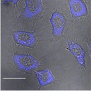
- Figure: Confocal fluorescence microscopic image merged with differential interference contrast image of living HeLa cells labeled with Pt5(MAA)8. The scale bar is 20 um.
[1] S. Tanaka, J. Miyazaki, D. K. Tiwari, T. Jin and Y. Inouye, Angew. Chem. Int. Ed. 50 431(2011).
1Department of Electronic Engineering, Ching Yun University, Jung Li, Taiwan
2 Instrument Technology Research Center, National Applied Research Laboratories, Hsinchu 300
3 Department of Physics, National Taiwan University, Taipei 10617
4 Department of Chemical Engineering, National Taiwan University, Taipei 10617
5 Research Center for Applied Sciences, Academia Sinica, Taipei 115-29
*E-mail: dptsai@phys.ntu.edu.tw
Enviromental applications of plasmonic effects for photo-catalytic chemical reactors will be discussed in this talk. Plasmonic hot spots and their near-field optical interactions are considered to be the novel methods to achieve high efficient photo-catalytic chemical process. The localized and enhanced electromagnetic field of plasmonic nanostructures of nano recording marks of optical disk provide many hot spots. We found the blinking experiments can demonstrate the hot spots of the optical disks used in our photo-catalytic reactor. Results of the photo-catalytic process happened in the plasmonic optical disk photo-chemical reactor successfully demonstrate better efficiency of the decomposition of the Methyl Orange to carbon dioxide and water.
[1] A. Fujishima & K. Honda, Nature 238, 37-38 (1972).
[2] M. R. Hoffmann, S. T. Martin, W. Choi, and D. W. Bahnemannt, Chem. Rev. 95, 69-96 (1995).
[3] R. Asahi, T. Morikawa, T. Ohwaki, K. Aoki, Y. Taga, Science 293, 269-271 (2001).
[4] A. L. Linsebigler, G. Lu, and J. T. Yates, Jr., Chem. Rev. 95, 735-758 (1995).
[5] I. M. Arabatzis, T. Stergiopoulos, D. Andreeva, S. Kitova, S. G. Neophytides, and P. Falaras, J. of Catalysis 220, 127-135 (2003).
[6] C. S. Wu, T. H. Wu, T. C. Chu, H. J. Huang, D. P. Tsai, "Application of Optical-fiber Photoreactor for CO2 photocatalytic Reduction," Topics in Catalysis 47, 131-136 (2008).
[7] Z. Y. Wang, H. C. Chou, C. S. Wu, D. P. Tsai, G. Mul,"CO2 photoreduction using NiO/InTaO4 in optical-fiber reactor for renewable energy," Applied Catalysis A: General 380 172–177 (2010).
1. Institut Fresnel, Aix-Marseille Université, CNRS, Ecole Centrale Marseille, Campus de St Jérôme, 13397 Marseille, France
2. Institut de Science et d'Ingénierie Supramoléculaires, Université de Strasbourg, CNRS, 8 allée G. Monge, 67000 Strasbourg, France
jerome.wenger@fresnel.fr
Controlling the fluorescence emission from nanoscale quantum emitters such as single molecules is a key element for a wide range of applications, from efficient analytical sensing to quantum information processing. Enhancing the fluorescence intensity and narrowing the emission directivity are both essential features to achieve a full control of fluorescence, yet this is rarely obtained simultaneously with optical nanoantennas.
Here we report that gold nanoapertures surrounded by periodic corrugations transform standard molecules into bright unidirectional fluorescence sources [1] (Fig. 1a & 1b). We obtain enhancement factors of the fluorescence rate per molecule up to 75 fold (Fig. 1c) simultaneously with a directional emission of the fluorescence into a narrow angular cone into a cone of ±15° in the direction normal to the sample plane (Fig. 1d). The bright emission and narrow directionality enable the detection of single molecules with a low numerical aperture objective, and improve the effectiveness of fluorescence-based applications.
We thoroughly quantify the increased light-matter coupling as well as the radiation pattern intensity. Our study also demonstrates efficient detection of single fluorescent molecules using a simple 0.5 NA water immersion objective, or equivalently a 0.4 NA dry air objective. This releases the need for high NA microscope objectives. Another remarkable point is that the structure is insensitive to the incoming polarization direction. We believe that the present demonstration is of high relevance for the development of advanced single molecule sensing, bright single-photon sources for quantum information processing, and light emitting devices.
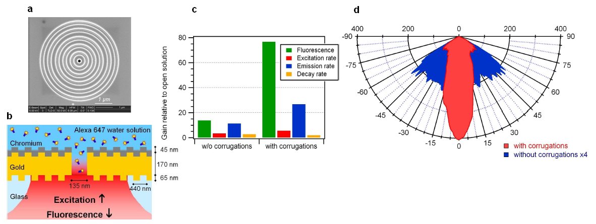
- Fig. 1 (a) Scanning electron microscopy image of the fabricated nanoaperture with five corrugations. (b) Configuration to enhance and control the fluorescence emission of molecules diffusing in the central aperture. (c) Contribution of excitation and emission gains to the overall fluorescence enhancement. (d) Angular radiation patterns in the polar angle, the intensity is calculated back to average signal per molecule.
[1] H. Aouani, O. Mahboub, N. Bonod, E. Devaux, E. Popov , H. Rigneault, T.W. Ebbesen, J. Wenger, "Bright unidirectional fluorescence emission of molecules in a nanoaperture with plasmonic corrugations", Nano Letters 11, 637-644 (2011).
Friedrich-Schiller-University Jena & Institute of Photonic Technology (IPHT) Jena
Structured illumination microscopy (SIM) is a well-established technique for the acquisition of confocal-like, optically sectioned images of biological specimen [1]. Its biggest potential however lies in high resolution imaging: illuminating a fluorescent sample with very fine light patterns will shift high spatial frequency information which cannot be detected by conventional wide-field microscopes into the support band of the microscope's optical transfer function (OTF) [2, 3]. The thus acquired information can be computationally shifted to its true positions in frequency space, effectively enlarging the OTF's support region and enhancing the microscope's resolution. In epi-fluorescence this approach can lead to a resolution improvement of about a factor two – or even higher, if nonlinear relations between illumination and emission intensity are exploited [4, 5].
Combining SIM with selective plane illumination microscopy (SPIM) yields the benefits of both techniques. This has been shown in the case of sectioning SIM [6]: here the excellent sectioning capability of SIM is enhanced by the fact that regions far out of focus are not illuminated and therefore no longer contribute to noise in the (computationally) sectioned images. Furthermore, illuminating only in-focus areas leads to a significant reduction of photo-bleaching in the whole sample.
But as for the epi-fluorescent case, the true potential of combining SIM and SPIM lies in high resolution imaging. While this combination benefits from the characteristic SPIM advantages of improved signal-to-noise levels and decreased photo-bleaching, selective plane illumination can furthermore help improve the advantages of SIM. Illuminating in a standing wave configuration under angles perpendicular to the optical axis allows for the finest possible grating illumination, and the resolution can be improved to beyond what would is possible in epi-mode, allowing an effective illumination numerical aperture of about 1.5.
We will give an introduction to SIM imaging and to the challenges associated with the reconstruction of high resolution images from the acquired data. Furthermore we will present strategies of how SIM and SPIM can be combined as well as simulations demonstrating the expected enhancement.
[1] M A A Neil, R Juškaitis, and T Wilson, Method of obtaining sectioning by using structured light in a conventional microscope, Opt. Lett. 22:1905-1907 (1997)
[2] R Heintzmann, and C Cremer, Laterally modulated excitation microscopy: improvement of resolution by using a diffraction grating, Proc. SPIE 3568:185-196 (1999)
[3] M G L Gustafsson, Surpassing the lateral resolution limit by a factor of two using structured illumination microscopy, J. Microsc. 198:82-87 (2000)
[4] R Heintzmann, T M Jovin, and C Cremer, Saturated patterned excitation microscopy – a concept for optical resolution improvement, J. Opt. Soc. Am. A 19:1599-1609 (2002)
[5] M G L Gustafsson, Nonlinear structured-illumination microscopy: Wide-field fluorescence imaging with theoretically unlimited resolution, PNAS 102:13081-13086 (2005)
[6] P J Keller, A D Schmidt, A Santella, K Khairy, Z Bao, J Wittbrodtm, and E H K Stelzer, Fast, high-contrast imaging of animal development with scanned light sheet–based structured-illumination microscopy, Nat. Meth. 7:637-642 (2010)
Institute of Chemistry, The Hebrew University of Jerusalem, Jerusalem 91904, Israel
willnea@vms.huji.ac.il
The coupling between the localized plasmon of Au nanoparticles (NPs) and the surface plasmon wave associated with Au-coated surfaces leads to the amplified detection of minute changes in the dielectric properties of surfaces. This is reflected by substantial shifts in the SPR spectra. Imprinted Au NPs composites on Au surfaces were prepared by the electropolymerization of thionaniline-functionalized Au NPs or ligand-modified thionaniline-functionalized Au NPs in the presence of a target substrate as imprint molecule. After removal of the imprint substrates, specific recognition sites for the association of the substrates are generated. These matrices are used as stereoselective or chiroselective sensor systems, using SPR as readout signals. Alternatively, the imprinted matrices act as selective "sponges" for the uptake and release of substrates. Imprinted matrices for the ultrasensitive selective detection of explosives (TNT, RDX, EGDN, PETN, nitroglycerin), for the stereoselective and chiroselective sensing of saccharides, amino acids, and antibiotics, and for the ultrasensitive detection of metal-ions were prepared.
Similarly, the imprinted Au NPs composites were implemented for the electrochemically or photochemically controlled uptake and release of substrates from the matrices.
Also, a series of amplified SPR sensors for DNA analysis, detection of aptamer-substrate complexes, or for the following of telomerase activity using Au NPs as amplifying labels will be discussed.
Finally, a new sensing platform that involves chemiluminescence resonance energy transfer (CRET) for bioanalytical applications will be introduced. The chemiluminescence generated by hemin/G-quadruplex structure is used as an internal light source for excitation of CdSe/ZnS quantum dots (QDs) and stimulation of the luminescence of the QDs. QDs are functionalized with nucleic acid nanostructures that include the caged sequence of the G-quadruplex. Upon detection of the respective DNA target or the formation of an aptamer-substrate complex, the hemin-G-quadruplexes are formed, resulting in the chemiluminescence of the QDs. Multiplexed analysis of DNA will be exemplified.
Institute of Photonic Technology, Albert-Einstein-Straße 9, 07745 Jena, Germany
*fritzsche@ipht-jena.de
Metal nanoparticles show not only exceptional electronic and optical properties but also a compatibility with the biomolecular world. Thus, they have attracted increasing attention with growing interest in several nanobiotechnological disciplines, such as the manipulation of biomolecules at the nanometer length scale. Here, we present the effects of excited silver nanoparticles attached to DNA molecules by fs laser pulses.
Introduction
Because of their unique optical properties based on localized surface plasmon resonance (LSPR), which can be tuned to specific wavelengths, e.g. by modifying the size, shape or material, noble metal nanoparticles can act as laser light antennas and allow a very local energy conversion of laser pulses from the nanoparticle into the surrounding medium. Thereby, the pulse width of the laser light plays an important role. While the excitation of metallic nanoparticles with nanosecond- or picosecond laser pulses results already during the pulse duration in an electron-phonon relaxation process and in an increased temperature in the proximal dielectric surrounding, resonant femtosecond laser pulses are too short and do not result in such a increase of the temperature [1]. However, highly excited electrons in the particles can easily overcome the metal's work function by absorption of three to four photons. These electrons and the amplified electric near field interact directly with the dielectric surrounding [2].
Results and Discussion
To study the interaction of femtosecond laser pulses with silver nanoparticle labelled DNA molecules, we used the UV- and electron sensitivity of an underlying Polymethylmethacrylate (PMMA) layer as an indirect detection method. First of all, we ensured that the DNA itself and the PMMA layer were not affected by femtosecond laser pulses. Afterwards, nanoparticle labelled DNA bundles were stretched onto the PMMA layer and irradiated with the same laser parameters, resulting in a quite impressive change of these DNA structures [3]. Thereby the plasmonic excitation caused by the irradiation of the nanoparticles is guided along the attached DNA-molecules, which results in a structuring of the PMMA along the former DNA position and leads to a thin groove reaching even micrometers away from the excited nanoparticle. These grooves are visible as dark structures in the AFM images with a depth of 3-4 nm. To explain this apparent energy transfer from the particle to the DNA, two mechanisms are eligible. On the one hand, the electrons of the nanoparticle could overcome their affinity and transfer their kinetic energy to the electrons of the PMMA and the DNA. On the other hand, a very high electric field (108-109 V/m) in the environment of the irradiated nanoparticle could accelerate electrons in the PMMA or DNA, which could result in a removal of PMMA side groups (radical formation) at a random position nearby the nanoparticle and the DNA, leading to a local PMMA degradation.
This combination of the optically controlled nanoantenna effect with the plethora of geometries provided by DNA could have applications in fields such as lithography, nanoplasmonics or molecular electronics.
[1] F. Garwe et.al., Nanotechnoloy 19, p.055207 (2008)
[2] A. Csaki et. al. Nano Lett. 7, p.247 (2007)
[3] J. Wirth et.al., Nano Lett., dx.doi.org:10.1021/nl104269x
1Institute of Physical Chemistry and Institute of Photonic Technology, Jena, Germany
2Institute of Photonic Technology, Jena, Germany
Photochemistry plays a keyrole in everyday life, e.g. life on earth relies on photosynthesis for oxygen generation and the production of carbohydrates. Currently research also focuses on understanding photocatalysis in order to exploit its potential for hydrogen production for the production of environmentally-friendly fuels. In this contribution novel artificial reaction centres have been synthesized based on photocatalytic active gold colloids and investigated by Raman spectroscopic techniques to localize the absorption process and study the electron transfer to the potentially catalytic gold colloid. Additionally, (DFT) calculations have been performed.
Successful conjugation of commercially available Ru-complexes (N749 and Z907, Solaronix) was determined by UV-vis spectroscopy. After conjugation the plasmon resonance shifts indicating effective binding of the dyes onto the surface. Subsequent surface enhanced Raman spectroscopy (SERS) of all three dye colloids definitely confirms binding by detecting their SERS spectrum at much lower concentrations than detectable by Resonance Raman spectroscopy, indicating SERS signal enhancement of at least three orders of magnitude. Furthermore quenching of dye luminescence and observation of Raman band shifts and changing band intensity ratios could be observed which are in good agreement with quantum chemical modelling.
The site of photoexcitation has been identified by comparing nonresonant FT-Raman and Resonance Raman spectra with quantum chemical calculations.
Financial support of the 'Thüringer Ministerium für Bildung Wissenschaft und Kultur (TMBWK)' of the project PhotoMIC (grant no.: B514-09049) is gratefully acknowledged. B.D. thanks the Fonds der Chemischen Industrie for financial support.
[1] M. Schmitt, S. Tschierlei, M. Karnahl, M. Presselt, C. Kuhnt, A. Yartsev, T. Pascher, V. Sundström, M. Schwalbe, B. Schäfer, S. Rau, B. Dietzek, and J. Popp, "Photophysics of an Intramolecular Hydrogen-Evolving Ru-Pd Photocatalyst," Chemistry - A European Journal, vol. 15, 2009, pp. 7678-7688.
[2] B. Dietzek, M. Schmitt, J. Popp, S. Tschierlei, M. Karnahl, M. Presselt, J. Guthmuller, L. González, and S. Rau, "Photochemisches Schicksal: Der erste Schritt bestimmt die Effizienz der H2-Bildung mit einem supramolekularen Photokatalysator," Angewandte Chemie, 2010, pp. n/a-n/a.
[3] C. R. Mayer, E. Dumas, F. Sécheresse, Journal of Colloid and Interface Science, 328 (2008), 452-457.