
JPK Instruments AG, Bouchestraße 12, Berlin, 12435, Germany, office@jpk.com
A trend in optical spectroscopy has emerged that makes use of the interaction of an AFM tip with a sample in an external optical field. Special attention has been devoted to tip enhanced Raman spectroscopy and to the manipulation of molecules and particles with simultaneous fluorescence. JPK picks up these trends and introduces the Tip Assisted Optics (TAO) module as an add-on to the NanoWizard AFM.
The NanoWizard AFM is designed for the integration with optical techniques on the basis of inverted optical microscopes. As a three-axis closed loop system it allows the sample to stay fixed within the optical focus. Furthermore, the use of an infrared light source (850nm) and special filters eliminates cross talk between optical experiment and AFM.
The TAO system consists of an additional 100x100µm X-Y-scanner for the sample, which is independently and simultaneously used with the AFM. This allows for the precise compensation of the offset between confocal image obtained with the sample scanner and the AFM topography. The tip can be positioned exactly into the optical focus while the user can navigate within the AFM image. Thus the tip-enhancement effect can be maximized and it becomes possible to do single molecule manipulation experiments within the focus of a confocal optical setup.
Max Planck Institut für Polymerforschung, Ackermannweg 10, D-55128 Mainz, Germany
Crescent-shaped structures have been prepared using a nanosphere lithographic (NSL) approach [1] and studied in terms of their optical response, in the visible and near infrared regimes. Geometrical parameters have been largely and systematically varied, and their influence on the multiple polarization dependant resonances studied [1,2]. A simple model based on that for resonances on metal stripes is presented as a first approach to describe this system [2,3].
The response of the crescents’ multiple resonances to a change in their dielectric environment has been investigated by attaching thin polyelectrolyte films to the structures. Based on these investigations, their environment sensing capabilities are presented and discussed.
Fluorescence enhancement on the structures prepared by colloidal lithography was investigated on a single object basis. Clear signatures of enhancement accompanying the resonances are present, both in single- and in two-photon excitation schemes.
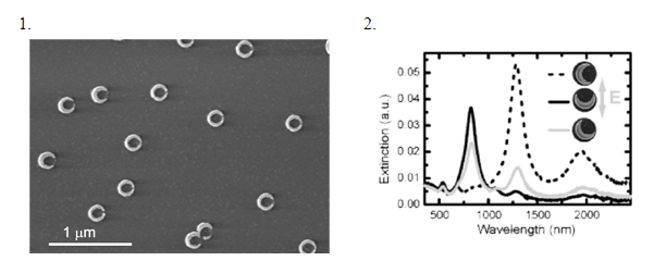
Fig. 1 Representative scanning electron micrograph of crescents prepared with via two consecutive metal depositions (Au, 2 x 20 nm, colloids’ diameter = 150 nm).
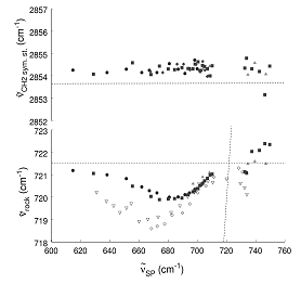
Fig 2. Polarized extinction spectra of gold crescents, prepared on a glass substrate (dcoll = 150 nm, 30 nm thick Au, 1 deposition)
[1] J. Shumaker-Parry, H. Rochholz, M. Kreiter, Advanced Materials 17, 2131–2134 (2005)
[2] H. Rochholz, N. Bocchio, M. Kreiter, New J. Phys. accepted (2007)
[3] J. Krenn et. al., Applied Physics Letters, 77, 21, 3379, (2000)
1JBCI, IPC/Friedrich-Schiller-University Jena, Helmholtzweg 4, D-07743 Jena, Germany
2IPHT Jena, Albert-Einstein-Straße 9, D-07745 Jena, Germany
For the analysis and investigations of inorganic and organic molecules Raman spectroscopy is a useful tool. However, the Raman effect is a very weak one thus affecting the detection limit. One possibility to increase the sensitivity is the enhancement of the Raman signals through an interaction between an analyte molecule and a roughened metallic surface. This technique, called Surface Enhanced Raman Spectroscopy – SERS allows a detection of molecules down to the single molecule level [1]. Traditional SERS active substrates are roughened metal electrodes, colloids, or metal layers produced by deposition. However, a common problem is the reproducibility of SERS substrates [2].
Within this contribution three different opportunities to produce reproducible SERS active substrates will be presented.
- Triangle nanostructures as SERS active substrates can be produced by nanosphere lithography – NSL [3]. Therefore a monolayer of polystyrene nanospheres in its closest package is deposit on a glass slide and covered with silver. After removal of the polystyrene beads nanotriangles are left.
- By electron beam lithography – EBL substrates with periodic diamond shaped structures are produced for analytics by means of SERS.
- Further silicon nanowires with gold caps [4] were tested as SERS active substrate. The nanowires can be produced by chemical vapour depostion on a gold implanted Si wafer.
The described SERS active substrates are tested with crystal violet for determination of the enhancement factor. In future work reproducible SERS active substrates will be used for detection of DNA and bacteria.
Acknowledgement
Funding of research project “Jenaer Biochip Initiative (JBCI)” within the framework “Unternehmen Region – Inno Profile” from the Federal Ministry of Education and Research, Germany (BMBF) is gratefully acknowledged.
[1] Kneipp K. et al., Physical Review Letters 1997, 78 (9), 1667.
[2] Vo-Dinh T. and Stokes D. L., SERS-based Raman Probes, in Handbook of Vibrational Spectroscopy (Wiley & Sons, Chichester) 2002, 2, Eds.: Chalmers, Griffiths, 1302.
[3] Haes A. J. et al., MRS Bulletin 2005, 30, 368.
[4] Stelzner et al., Nanotechnology 2006, 17, 2895.
The Ohio State University, 100 W. 18th Ave., Columbus, OH 43210-1173, USA
The surface plasmon (SP) mediated, extraordinary transmission of metal arrays of subwavelength holes has been moved into the infrared (IR) region in order to overlap with the traditional range of molecular vibrations. Enhanced IR absorption spectra are recorded[1] (using standard FTIR instrumentation) of metal-supported self-assembled alkanethiol monolayers[2], phospholipid bilayers[3], gramicidin[4] (an antibiotic peptide) and cholesterol in phospholipid bilayers, as well as hexadecane thin films. The interaction of a SP resonance and a vibrational excited state has been examined by tuning a SP resonance (both by film thickness and angle of the mesh) through the primary rocking vibration of the hexadecane molecule producing vibrational band intensity changes, peak shifts, and lineshape changes. The nature of the enhancements will be discussed.
The frequency of two hexadecane vibrations [CH2 symmetric stretch (top, not resonant) and CH2 rock (bottom, resonant)] as a function of the position of the (1,0)- SP transmission resonance. The CH2 rocking vibration shows systematic shifts in response to the SP. A mesh with a stronger SP transmission resonance (lighter symbols in bottom plot) shows a larger shift in frequency. Shifts this size in vibrational Stark spectroscopy require field strengths on the order of MV/cm.
[1] J.V. Coe et al., Analytical Chemistry, Feature Article, March 1, 78(5), 1385-1390 (2006).
[2] K.R. Rodriguez et al., J. Chem. Phys. 121(18), 8671-8675 (2004).
[3] S.M. Teeters-Kennedy, et al., J. Phys. Chem. B, 110(43) 21719-21727 (2006).
[4] J.V. Coe et al., Nanotechnology, 15 S495-S503 (2004).
Institute for Photonic Technology Jena
The plasmonic properties of nanoscale metal structures depend on parameters such as material composition, dimensions, shape and optical properties of the surrounding media. Changes in these parameters can be detected by measurements of the spectral shift of the plasmon absorption band. Based on optical darkfield detection of metal nanoparticles [1], we present results following changes in size, composition or surrounding media on a single particle level and in combination with other (e.g. AFM) characterization techniques. On the other hand, designer particles with a chosen absorption band are possible by e.g. choice of the material composition in the case of core-shell particles [2].
In order to access these effects for applications e.g. in bioanalytics or other sensoric use, time- and cost-effective methods to follow the spectral changes are required. One possibility is the in-situ monitoring of particles based on color changes, that can be transformed (using calibration curves) into e.g. particle dimensions for known material composition and shape. Other applications of the plasmonic properties of metal nanoparticles include the use of particles as nanoantennas, enabling optical manipulation of e.g. genetic material in a parallel approach and with sub-wavelength precision that is mainly determined by the size of the nanoparticles. Experimental results with human metaphase chromosomes demonstrate the potential of this approach [3].
[1] H. Siedentopf and R. Zsigmondy, Annalen der Physik 315 (1902) 1.
[2] A. Steinbrück, A. Csaki, G. Festag and W. Fritzsche, Plasmonics 1 (2006) 79.
[3] A. Csaki, F. Garwe, A. Steinbrück, G. Maubach, G. Festag, A. Weise, I. Riemann, K. König and W. Fritzsche, Nano Letters 7 (2007) 247.
Universidad Carlos III de Madrid (UCIIIM)
W. Fritzsche, A. Csaki, G. Festag, A. Steinbrück
Institut für Physikalische Hochtechnologie (IPHT)
Use of Gold Nanoparticles (NPs) as a contrast agent is shown to improve the efficiency of photoacoustic signal generation [1], thus enhancing the contrast between two different tissue types. In this paper Gold NPs are investigated for the detection of abnormalities in human soft tissue. The use of Gold NPs in the biomedical industry is already achieving great success. Oraevsky, A.A., has shown that it is possible to differentiate between malignant and benign tumours in breast tissue using the photoacoustic technique, and has increased the contrast 2 - 6 fold when using Gold NPs[2]. Besides this Zharov, V.P. et al have used gold NPs conjugated with specific antibodies for the selective killing of bacteria using photothermal nanotherapeutics[3].
In this paper an analysis of the absorption properties of Gold NPs will be investigated for photoacoustic signal generation. This study compares the signals produced from NPs of different geometrical characteristics. To perform this test the NPs will be inserted in a gel matrix which is used as a phantom to mimic soft tissue properties. Using the Time of Flight (TOF) analysis the localization of the Gold NPs within the gel phantom is obtained. Using this technique and an experimental scheme consisting of an acoustic transducer which rotates around the phantom has given a two dimensional image of the nanoparticle source in the phantom. This work is part of an ongoing process for the development of a low cost 3D imaging scheme for cancer detection.
This work belongs to IPHT (Germany) and UCIIIM (Spain) under the Integrated Action Collaboration on “Investigation of Optical Diffused and Optoacoustic Breast Cancer Detection by using High Contrast Nanoparticles Agents ” (2006 – 2007).
[1] Copland J.A., Eghtedari M., Popov V.L., Kotov N., Mamedova N., Motamedi M., Oraevsky A.A. “Bioconjugated gold nanoparticles as a molecular based contrast agent: implications for imaging of deep tumors using optoacoustic tomography”, Molecular Imaging Biology, 6(5), pages 341-9, Sept-Oct 2004.
[1] Oraevsky, A.A., “Detection, diagnostics and image-guided therapy of cancer using laser optoacoustic imaging system and gold nanoparticles”, Biophotonics, 2004. APBP 2004. The Second Asian and Pacific Rim Symposium, pp 250 – 251, 14 17 December 2004.
[3] Zharov, V.P., Mercer, K.E., Galitovskaya, E.N., Smeltzer, M.S., “Photothermal Nanotherapeutics and Nanodiagnostics for Selective Killing of Bacteria Targeted with Gold Nanoparticles”, Biophysics Journal, Vol. 90., pp 619 – 627, January 2006.
SUPA, School of Physics and Astronomy, University of St. Andrews, North Haugh, St. Andrews, Fife, KY16 9SS, United Kingdom
Nano-sized metal particles have become a remarkable tool for various applications in nanotechnology. They may enhance fluorescence [1] as well as offering an extremely precise instrument for molecular surgery on chromosomes [2]. In addition, trapping colloidal silver particles opens up new possibilities for surface-enhanced Raman spectroscopy (SERS) [3].
An intriguing property of metal nanoparticles is their intrinsic plasmon resonance, depending upon size, shape and material of the particle. This resonance is the underlying mechanism of their ability to enhance and localise electromagnetic fields in their close surrounding. The plasmon resonance results in a large polarisability of metal nanoparticles thus an increased dipole force favourable for optical trapping. It is now well established that a wide range of differently sized metal nanoparticles can be trapped in conventional optical tweezers [4]. Until now experiments to trap gold spheres are carried out far (to the red) from resonance mainly at 1064nm. In that regime the dipole force is attractive for the metal particles. The far red-detuning is required to reduce heating effects that are an issue, increasing the Brownian motion of the particle [5].
We present to our knowledge the first blue-detuned optical trap for gold nanospheres [6]. This exploits the plasmon resonance such that the trapping wavelength is below the resonance of the particles, where they experience a repulsive dipole force. Such a trap requires optical beamshaping in a way that we may confine the nanoparticles to a dark region. To achieve this we use an annulus of light surrounding the gold nanosphere preventing it from escaping the trap as it gets repelled by the light around it. Laguerre-Gaussian (LG) laser beams offer the advantage of such a dark core (vortex), whose size depends upon the topological charge l [7]. We investigate the wavelength dependence to stabilize gold particles in the vortex of an LG beam with respect to the power required. By using different sizes of LG beams (different topological charge l) the area of confinement can be controlled and the amount of particles taking up at once inside the trap is adjustable. The advantage of confinement is that particles remain in the dark region hence experiencing less scattering events avoiding strong absorption and heating.
[1] S. Kühn et al., Physical Review Letters 97, (2006), 017402
[2] A. Csaki et al., Nano Letters 7, (2007), 247
[3] F.Svedberg et al., Nano Letters 6, (2006), 2639
[4] P.M. Hansen et al., Nano Letters 5, (2005), 1937
[5] Y.Seol et al., Optics Letters 31, (2006), 2429
[6] M. Dienerowitz et al., to be submitted
[7] M. A. Clifford et al., Optics Communications, 156, (1998), 300
Institute for Photonic Technology Jena
The plasmonic properties of nanoscale metal structures depend on parameters such as material composition, dimensions, shape and optical properties of the surrounding media. Deposition of metal shells on e.g. gold nanoparticles by reductive metal deposition as known from electron microscopy allows for the defined and highly specific growth of the seed particles. We followed this growth on a single particle level, imaging the same set of particles after every step of metal deposition [1]. In combination with single particle spectroscopic characterization, this technique allows for a correlation of size and composition with the plasmonic properties. We extended this method to the investigation of enzymatic systems for nanoscale metal deposition that lead to highly anisotropic metal structures [2].
[1] G. Festag, A. Steinbrück, A. Csaki, R. Möller and W. Fritzsche, Nanotechnology 18 (2007) 015502.
[2] R. Möller, R. D. Powell, J. F. Hainfeld and W. Fritzsche, Nano Letters 5 (2005) 1475.
Heinrich-Heine-Universität Düsseldorf, Institut für Physicalische Chemie, Düsseldorf, Germany
Well studied mechanical properties of DNA molecules, and the ability to create DNA constructs having various sequence and modifications, provide an excellent test sample for new experimental technique establishment. At the same time, important role of DNA molecules in cells replication, protein building, etc. makes them an attractable object for scientific research.
Here we present results on new technique developed in our lab: simultaneous atomic force and fluorescence spectroscopy (SFFS) [1]. In one of experimental approaches fluorescence or scattering signal from a sample is used for the force spectroscopy spatial localization. After the sample molecule localization multiparameter fluorescence detection (MFD) allows one to obtain information about photophysical properties of fluorescence emitters and conformational changes of macromolecules [2,3]. It was also recently demonstrated that probability distribution analysis (PDA) can extract intermolecular distance from fluorescence resonance energy transfer (FRET) measurements with sub-angstrom precision [4]. Mechanical manipulation of the sample adsorbed on the surface increases the amount of information accessible, however putting experimental limitations too [5].
The scope of the SFFS technique is demonstrated in studies of λ-digest DNA molecules and DNA molecules modified for specific binding. SYBR Green I fluorescent dye influence is investigated by force spectroscopy. Simultaneous fluorescence and atomic force spectroscopy measurements will be presented.
[1] Gaiduk A., et al., ChemPhysChem., 2005, 5, 976.
[2] Eggeling C., et al., J.Biotechnology, 2001, 86, 163.
[3] Rothwell, et al., PNAS, 2003, 100, 1655.
[4] Antonik M., et al., JPCB, 2006, 110, 6970.
[5] Gaiduk A., et al., MRT, 2007, 70, 433.
International Centre of Biodynamics, 1 B Intrarea Portocalelor, 060101, Bucharest 6, Romania, www.biodyn.ro, mgheorghiu@biodyn.ro
Surface functionalization and nanopatterning ad the required selectivity and specificity to the otherwise nonspecific, yet powerful methods like electrochemical and surface plasmon resonance (SPR) assays for biosensing. In the same time, it can provide signal enhancement, background reduction and controlled interaction with biological systems.
We will present some of our latest developments on harvesting the enhancing capabilities of nanogold (thin coating and nanobeads) plasmonics in conjunction with affinity compounds for biosensing: biosensing platforms development, modeling, data analysis and combined (electrochemical and optical) analysis set-ups, evolving to open new combinations of methods with improved technical performance, helping to resolve challenging bioanalytical problems including sensitivity, signal resolution and specificity by interfacing these technologies in small volumes. Methods Multichannel, differential impedance spectroscopy either alone or in combination with complementary methods: surface plasmon resonance (SPR), fluorescence microscopy, electrochemistry to develop, optimize and analyze specific nanostructured platforms (affine and cell based biosensors).
Selected results dealing with detection of target compounds and pathogens, surface interaction / and cell adhesion control through nanopatterning will be presented. The capabilities of combined electrooptical methods to study nanostructured biointerfaces and possible applications in life sciences will be outlined.
University of California San Diego, Department of Bioengineering and Department of Electrical and Computer Engineering, La Jolla, CA 92093-0412 USA
Despite the advent of individually detectable nanoparticles, single molecule resolution has yet to translate into a viable platform for mapping the single base genetic changes associated with genetic disease and early stages of cancer. Sensitive assays are generally compromised when converted to complex biological samples such as tissue sections, smears, or whole blood. In particular, nanoparticle probes meant to detect DNA by hybridization regularly bind to cell matrices, proteins and other sections of the genome. For rare (low copy number) targets, this background is often the lower limit of detection. We have found that inherent bistabilities within DNA assembled fluorescent resonant energy transfer systems demonstrated time-varying optical signals in response to an electrophoretic driving force. Frequency responses of electrophoretically driven FRET systems were shown to be DNA sequence specific. Integration of these signals over time gave near single molecule sensitivity within a high background of autofluorescence. This research suggests that externally driven nanoscale mechanical systems may help improve information flow within morphologically intact specimens. We are further exploring both electrophoretic and acoustical perturbation of DNA FRET constructs and plasmonic nanoparticle constructs as a way to carry out single nucleotide polymorphism detection in un-amplified DNA samples.
Michael J. Heller received his Ph.D. in Biochemistry from Colorado State University in 1973. He was an NIH Postdoctoral Fellow at Northwestern University from 1973 to 1976. Dr. Heller was supervisor of the DNA Technology Group at Amoco Corporation from 1976 to 1984, and then the Director of Molecular Biology at Molecular Biosystems, Inc., from 1984 to 1987. Dr. Heller was a co-founder of Integrated DNA Technologies, and served as President and Chief Operating Officer from 1987 to 1989. He was a co-found of Nanotronics and Nanogen, and served as the Chief Technical Officer of Nanogen from 1993 to 2001. Dr. Heller is now a professor in the departments of Bioengineering and Electrical and Computer Engineering at the University California San Diego, and still serves as a consultant to Nanogen. Dr. Heller has extensive industrial experience in biotechnology, biomedical devices and nanotechnology. Dr. Heller has a respectable publication record, and has been an invited speaker to a large number of scientific conferences and meetings. He has over 35 issued US patents, including some key patents on self-assembly nanofabrication. Dr. Heller has been a panel member for the White House (OSTP) National Nanotechnology Initiative 1999/2000; the NAS (NAE) Review of National Nanotechnology Initiative 2001-2002; the NAS(NAE) – Engineer for the 2020 - 2001/2002; and has been involved in a number of NSF Nanotechnology Workshops. He is now a panel member for the California Blue Ribbon Task Force on Nanotechnology.
1 JBCI, Institute of Physical Chemistry; Friedrich-Schiller-University Jena, Helmholtzweg 4, D-07743 Jena, Germany
2 Institute of Photonic Technology, Albert-Einstein-Straße 9, D-07745 Jena, Germany; E-Mail: Juergen.Popp@uni-jena.de
Over the past years many detection techniques for DNA sequences were developed. The aim was to overcome the disadvantages of the established method, fluorescence spectroscopy. One greatly promising technique is Surface Enhanced Resonance Raman Scattering (SERRS). In comparison to fluorescence, SERRS can provide at least nearly equal detection limits [1]. Additionally, by exploitation of the narrow-band spectroscopic fingerprints of the Raman-active dyes multiplex identification of specific sequences of DNA is easily practicable [2,3,4].
One requirement for the enhancement of the Raman signal is the need of molecules with chromophor systems, which have an absorption maximum close to the laser frequency. Additionally the dye must be very close or in contact with a rough metal surface.
In this study short synthetic oligonucleotides, which are immobilised on glass surfaces are used. A new option of linking the label to the DNA sequence is tested in combination with the possibility of bringing the silver nanoparticle in close proximity to the dye by enzymatic silver deposition. As Raman active label commercially available fluorescence dyes are used.
Acknowledgement
The Funding of the research project “Jenaer Biochip Initiative (JBCI)” within the framework “InnoProfile - Unternehmen Region” from the Federal Ministry of Education and Research (BMBF) Germany is gratefully acknowledged.
[1] K. Faulds, R.P. Barbagallo, J.T. Keer, W.E. Smith, D. Graham, Analyst 129, 567-568 (2004)
[2] K. Faulds, W.E. Smith, D. Graham, Anal. Chem. 76, 412-417 (2004)
[3] D. Graham, B.J. Mallinder, W.E.Smith, Biopolymers (Biospectroscopy) 57, 85-91 (2000)
[4] Y.C. Cao, R. Jin, C. Mirkin, Science 297, 1536-1540 (2002)
1Walter Schottky Institute, Technical University Munich, Garching, Germany
2University of Leicester, UK
3Ludwig-Maximilians-University Munich, Germany
The growth of metallic nanoparticles along synthetic DNA templates constitutes a highly promising new route to yield nanoparticles of well controlled size and shape. Further, taking advantage of the superior recognition characteristics of nucleic acids, it should be possible to arrange DNA-based nanoparticles in self-assembled structures with defined inter-particle spacing.
In this study, we use two different methods to deposit silver on oligonucleotides of different lengths (23-96 base pairs) in order to synthesize metallic nanorods with controlled aspect ratios. The first method involves the specific labeling of nucleotides with aldehyde groups, followed by the exposure to Tollens reagent and a development solution, whereas the second method relies on the photo-induced deposition of Ag onto unmodified DNA samples. Several preparation parameters (DNA sequence, buffer salt type, Ag concentration, UV illumination time) are varied systematically.
The obtained DNA-templated nanoparticles are characterized with respect to their optical and structural properties. Plasmonic modes can be identified from maxima observed in the extinction spectra. The size and shape of the nanoparticles is inferred from conducting polarized dynamic-light-scattering experiments in the solution phase, as well as from imaging dispersed nanoparticles on single-crystalline substrates by atomic force microscopy.
1Experimentalphysik I and Center for Interdisciplinary Nanostructure Science and Technology – CINSaT, Universität Kassel, Heinrich-Plett-Str. 40, 34132 Kassel, Germany
2Institute of Optics, Technical University Berlin, Hardenbergstr. 36, 10623 Berlin, Germany
In this contribution we present surface-enhanced Raman scattering (SERS) measurements of Pyrene obtained with tailormade silver nanoparticles on dielectric substrates. The tailoring of the SERS substrates have been achieved by precise tuning of the localized surface plasmon polariton resonance of silver nanoparticles to the vicinity of the laser wavelength used for SERS excitation. The underlying method relies on control of the growth kinetics of supported metal nanoparticles which causes a pronounced shape change as a function of particle size. With this preparation method, SERS substrates with optimized plasmon resonances and field enhancement can be easily produced for specific excitation wavelengths and detection ranges.
As a first proof of the usefulness of our SERS-substrates, three silver nanoparticle ensembles with aplamon resonance at about 2.18 eV (570 nm), and one ensemble with a resonance at about 2.56 eV (485 nm) were prepared with and without protective coatings consisting of CaF2 (cf. Tab. 1). Subsequently, Raman-spectra of pyrene were recorded with an excitation wave-length of 514.5 nm (2.41 eV). For all nanoparticle ensembles, similar SERS spectra were ob-tained, indicating the high reproducibility of the substrates (Fig. 1). A detailed investigation revealed that the signal-to-noise ratio is significantly smaller for substrate B as compared to the other ones. The reason is the slightly detuned plamon resonance of the nanoparticles at 2.56 eV. Thus, fewer nanoparticles are in resonance with the laser frequency compared to substrates A, C and D. This result demonstrates clearly, how crucial the Raman-signal depends on the morphology of the nanoparticle ensembles and how important it is to use tailor-made nanoparticles to obtain optimum Raman-signals.
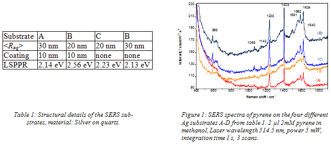
Interestingly, the ensembles coated with a 10 nm thick CaF2 protective layer exhibited still 70% of the enhancement compared to the bare nanoparticles. This is quite promising because protective coatings are essential for applications under a variety of conditions especially when long-term stability is required. Another important point is that all four investigated nanoparticle ensembles were very stable during the measurements, an advantage often not found for other substrates, although they may exhibit higher enhancement factors. Further tailoring of nanoparticles by laser irradiation is in progress to develop substantially improved SERS sub-strates. In summary, supported tailor-made nanoparticles produced by Volmer-Weber-growth are very promising candidates for new SERS-substrates.
Dep. of Applied Physics Chalmers University of Technology 412 96 Göteborg, Sweden kall@fy.chalmers.se
In this presentation, I will discuss some of our recent work on localized plasmons in gold and silver nanostructures, focusing on the importance of electromagnetic coupling between plasmon modes in strongly interacting systems. This phenomenon turns out to be crucial for a wide range of applications, including surface-enhanced spectroscopy, refractive index sensing and metamaterials, as illustrated in Figure 1-3.
Figure 1. Coupling between plasmons on the inner and outer surfaces of gold nanorings results in a hybridised dipolar mode in the NIR that exhibit promising characteristics for refractive index sensing of macromolecule biorecognition reactions. Reproduced from J. Aizpurua et al., PRL 90, # 057401 (2003) (left) and E. Larsson et. Al, Nano Letters (2007) in press
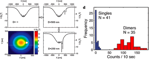
Figure 2. Near-field coupling between colloidial Ag particles captured by an optical tweezers results in an interparticle optical force that greatly amplify the SERS cross section from adsorbed benzenethiol. Reproduced from F. Svedberg et al., F. Svedberg et al., Nano Lett. 6, 2639-2641 (2006).
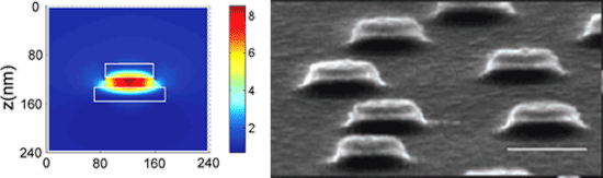
Figure 3. Coupling between the upper and lower Au disks in Au-SiO2-Au nano-sandwiches results in a hybrididized plasmon mode that strongly enhance the local magnetic field. This may be a route to metamaterials that exhibit negative refraction in the visible. Reproduced from T. Pakizeh et al, Optics Exp. 14, 8240-8246 (2006), A. Dmitriev et al., Small 3, 294-299 (2007).
1Harvard Medical School, Wellman Center for Photomedicine, Boston, MA
2Federal Institute for Materials Research and Testing, Berlin
3Harvard-MIT Division of Health Sciences & Technology, Cambridge, MA
Two-photon excitation is gaining rapidly in interest and significance in spectroscopy and microscopy.
We introduce a new approach that suggests versatile optical labels suitable for two-photon excitation and also two-photon excited ultrasensitive, non-destructive chemical probing. The underlying spectroscopic effect is the incoherent inelastic scattering of two photons on the vibrational quantum states called hyper Raman scattering (HRS). HRS results in Raman signals shifted relative to the second harmonic of the excitation laser. The effect is extremely weak. Cross sections on the order of 10 65 cm4s/photon (35 orders of magnitude smaller than those for Raman scattering !) make the use of hyper Raman scattering as a practical spectroscopic tool nearly impossible.
However, the rather weak effect can be greatly strengthened if HRS takes place in the local optical fields of gold and silver nanostructures. This so-called surface enhanced hyper Raman scattering (SEHRS) is the two-photon analogue to surface enhanced Raman scattering (SERS). SEHRS provides structurally sensitive vibrational information complementary to those obtained by SERS. The effect combines the advantages of two-photon spectroscopy with the structural information of vibrational spectroscopy, and the high sensitivity and nanometer-scale local confinement of plasmonics-based spectroscopy.
We show that two-photon cross sections for SEHRS can be on the order of 104 - 105 GM, and discuss spectroscopic applications of the effect.
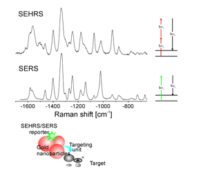
The Figure shows the concept of an optical labels suitable for 1- and 2-photon excitation based on SEHRS and SERS signals of a reporter molecule linked to a gold nanoaggregate: As an example, SEHRS and SERS spectra of Rose Bengal as reporter are displayed.
Biosensors Group. Microelectronics National Centre (IMM-CNM-CSIC). Isaac Newton 8, Tres Cantos, Madrid, 28760, Spain. Laura@imm.cnm.csic.es
SPR biosensing has focused a strong effort since the technique was first applied in 1983 and has become one of the most successful label-free and commercially accepted optical biosensor1. This technique has been employed in biomolecular engineering, drug design, monoclonal antibody characterization, epitope mapping, phage display libraries, virus-protein interaction, environmental pollutants detection, among other interesting problems.
We have developed a fully-automated biosensor SPR biosensor that uses a label-free format, (β-SPR from Sensia S.L., Spain2). This device has two flow cells of 300 nL and integrated control of pumps and valves for sample injection. The system is portable and incorporates software for data acquisition and instrument control. We will show the applicability of the device in the following fields:
- Our sensor is the first SPR-based immunosensor applied to the detection and validation of three relevant pesticides (DDT, chlorpyrifos and carbaryl) with higher sensitivity than other immunochemical systems (e.g. ELISA). Measurements of the pollutants are carried out using inhibition binding assays with sensitivity at ppt level. Although the SPR system has only two channels, we have implemented a new procedure for simultaneous multi-analyte determination. Environmental real water samples has been analysed and we have demonstrated that our level of detection accomplishes the EU legislation.
- The SPR sensor can also be used as an on-line immunoanalytical method for the evaluation of pesticide metabolites which can be present in the human body. We have detected the metabolite from chlorpyrifos pesticide (3,5,6-trichloro-2-Pyridinol (TCP)) from its primary via of elimination (urine). The immunoassay format allows a highly sensitive detection of TCP in human urine without the need of previous clean-up and preparation of samples. The comparison between TCP limits of detection in urine and assay buffer used as control showed similar sensitivity values and a limit of detection of 0.1 µg L-1 was obtained for TCP urinary determinations.
- The SPR technique has been also applied to the real-time and label-free detection of DNA hybridization and single point mutations. We have studied the mutations at gene BRCA-1 related to the predisposition in women to develop an inherited breast cancer. The SPR sensor has successfully discriminated between normal and mutant sequences with a limit of detection of only 100 nM. The device could be employed for the analysis of point mutations in the DNA samples of patients.
For many applications, improvement in the sensitivity of the SPR sensors is an important issue, and we are investigating several alternative configurations as Localised Surface Plasmon Resonance (LSPR) based on nanoparticles or Magnetooptical Surface Plasmon Resonance sensor (MOSPR) based on nanoestructures. The versatility, tunable properties, sensitivity and multiplexing capabilities of metallic nanostructures turn them into very qualified candidates to develop commercial biosensors in the near future.
(1) Lechuga LM (2005) In: Gorton L (ed) Biosensors and modern biospecific analytical techniques. Comprehensive analytical chemistry series, vol XLIV. Elsevier, Amsterdam, The Netherlands, chap 5
(2) www.sensia.es
(3) E. Mauriz, A. Calle, A. Abad, A. Montoya, A. Hildebrandt, D. Barceló and L.M. Lechuga. (2006) Biosens. Bioelectron. 21(11).
Institute of Physics and Erwin Schrödinger Institute for Nanoscale Research, Karl-Franzens University, 8010 Graz, Austria
We investigate the modifications of the spontaneous emission process in a europium chelate dye by coupling to localized plasmons in nanoparticles. The nanoparticles which are fabricated by electron beam lithography are arranged in a 2D lattice on a transparent substrate. The fluorophore-plasmon coupling can increase radiative and non-radiative de-excitation rates by orders of magnitude with respect to the unperturbed case [1]: When the plasmon resonance overlaps well with the molecules’ fluorescence emission frequency, the interacting molecules show a dramatically shortened fluorescence lifetime but an enhanced signal amplitude, as is evident in Fig. 1.
This mechanism sensitively depends on the mutual distance of fluorophore and particle which makes it a promising tool for new labeling techniques in biosciences and for modifications of organic light emitting diode emission properties.
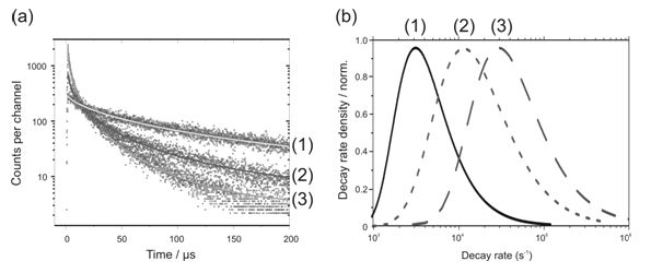
Figure 1: Time-resolved intensity decay curves (a) of the fluorophores on a bare silica substrate (1) and a non-resonant (2) and resonant (3) nanoparticle array. (b) shows the corresponding decay rate distributions.
[1] S. Gerber, F. Reil, U. Hohenester, T. Schlagenhaufen, J. R. Krenn and A. Leitner, Phys. Rev. B.. 75, 073404 (2007).
1Centre for Photonics and Photonic Materials, Department of Physics, University of Bath, Bath, UK
2Departamento de Física Teórica de la Materia Condensada, Universidad Autónoma de Madrid, Madrid, Spain
3Departamento de Física de la Materia Condensada, Universidad de Zaragoza, Zaragoza, Spain
The confinement of electromagnetic energy to volumes below the diffraction limit leads to a concomitant field enhancement, enabling a wealth of opportunities for sensing and spectroscopy. One of the challenges is to create such confinement in a controlled manner, and to channel electromagnetic energy from conventional, wavelength-scale devices such as planar waveguides and optical fibres down to the nanoscale. The field of plasmonics holds the promise of achieving this via the excitation of electromagnetic surface modes at the interface between a conductor and a dielectric. Depending on the exact geometry and the frequency of the electromagnetic radiation, a deep sub-wavelength mode size is possible [1, 2].
In the first part of the talk, we will present results for the controlled focusing of electromagnetic radiation at frequencies from the visible to the THz regime to sub-wavelength volumes, with a view to applications in biological sensing. While for visible and near-infrared frequencies conical metallic tips [3] coupled to conventional optical waveguides are a convenient means for nanoscale focusing, at lower frequencies the situation is more challenging. Here, the frequency of excitation is far below the intrinsic plasma frequencies of metallic conductors, and surface plasmon polaritons acquire the character of delocalized Sommerfeld-Zenneck waves. Sub-wavelength energy localization can however be achieved periodic surface structuring, leading to designer electromagnetic surface modes termed spoof surface plasmon polaritons [4]. While structured planar surfaces enable a new infrastructure for THz technology [5], corrugated cylindrical and conical structures show a high promise for the guiding and super-focusing of THz radiation (Figure) [6]. Analytical and numerical electromagnetic modelling of such structures will be discussed, and first implementations be presented.
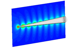
Figure: Focusing of electromagnetic radiation at 0.6 THz on a corrugated conical taper via spoof surface plasmon polaritons
In the second part of this talk, we will discuss various approaches for confining electromagnetic energy at visible and near-infrared frequencies using hybridized modes in designer cavities consisting of planar metallic structures with nanoscale gaps. Results from finite difference time domain simulation and near-field optical spectroscopy will be presented, and implications for biosensing discussed.
[1] Ozbay, E., Plasmonics: Merging photonics and electronics at nanoscale dimensions. Science, 2006. 311: p. 189.
[2] Maier, S.A. and H.A. Atwater, Plasmonics: Localization and guiding of electromagnetic energy in metal/dielectric structures. Journal of Applied Physics, 2005. 98: p. 011101.
[3] Stockman, M.I., Nanofocusing of optical energy in tapered plasmonic waveguides. Physical Review Letters, 2004. 93(13): p. 137404.
[4] Pendry, J.B., L. Martin-Moreno, and F.J. Garcia-Vidal, Mimicking surrface plasmons with structured surfaces. Science, 2004. 305(5685): p. 847.
[5] Maier, S.A. and S.R. Andrews, Terahertz pulse propagation using plasmon-polariton-like surface modes on structured conductive surfaces. Applied Physics Letters, 2006. 88: p. 251120.
[6] Maier, S.A., et al., Terahertz surface plasmon polariton propagation and focusing on periodically corrugated metal wires. Physical Review Letters, 2006. 97: p. 176805.
1School of Chemistry, Raymond and Beverly Sackler Faculty of Exact Sciences
2Department of Biochemistry, George S. Wise Faculty of Life Sciences, Tel Aviv University, Tel Aviv, 69978, Israel
The work to be presented has two aspects:
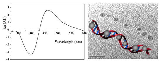
- The influence of chiral template moleculaes on the plasmons of metal nanoparticles[1]: Silver nanocrystals were grown directly on a poly(dG)-poly(dC) double stranded DNA scaffold by complexing the DNA with Ag+ and reducing the silver ions. The CD spectrum of the DNA-Ag nanocrystal complexes displayed a band centered at 425 nm which corresponds to the surface plasmon excitation of the nanocrystals. This chiral plasmon signature was not observed in a control experiment where silver nanocrystals of similar size were produced without the DNA template and adsorbed to the DNA. Thus, it was concluded that the DNA template induced the growth of asymmetric silver nanocrystals with a favored handedness.
- The influence of plasmons of metal nanoparticles on the spectroscopy of chiral molecules. Preliminary results on this subject will be presented.
[1] G. Shemer, O. Krichevski, G. Markovich, T. Molotsky, I. Lubitz, A. B. Kotlyar, "Chirality of Silver Nanoparticles Synthesized on DNA", J. Am. Chem. Soc. 128, 11006 (2006).
1Belarusian institute of system analysis, Department of innovation research, pr. Pobediteley 7, Minsk, 220004, Belarus
Philips Classic Laser Laboratories, University of Arkansas for Medical Sciences, Little Rock, Arkansas, 72205, USA
Gold nanoparticles (GNs) can be used for thermal mechanisms of selective nanophotothermolysis of cells under action of short laser pulses. Mechanisms of selective nanophotothermolysis with GNs with sizes 10-100 nm and short laser pulses with duration 10–6 <= tp <= 10–12 s were theoretically investigated. Modeling of mechanisms of selective nanophotothermolysis of cells with short laser pulses and gold nanoparticles are performed with focus on the thermal protein denaturation, photoacoustic effect due to the thermal expansion of nanoparticles, and explosive vaporization of liquid around the nanoparticle accompanied to acoustic and shock waves.
A simple analytical model for heating of an absorbing nanoparticle in tissue by a laser pulse was developed. Heating of the bulk (or of the surface) of a particle can initiate such processes as thermal denaturation of tissue proteins around GNs, thermal expansion of GNs and explosive formation of vapor in the water-contained tissues. The model can be used for description of the temperature rise in absorbing nanoparticles under specific experimental conditions and the likelihood of the occurrence of a specific physicochemical process that determines the final results (coagulation, disruption) of the laser pulse action on cells and tissues. Thermal radiation of bioconjugated and heated nanoparticles can be used for diagnostics of different targets inside tissues such as tumor cells.
Irradiation of GNs with sizes 10-100 nm by nano-, pico- and femtosecond laser pulses with different wavelengths could result in thermal and (or) mechanical intracellular effects with spatial confinement of about or less than 10-100 nm. Characteristic parameters of these processes such as the temperatures levels, thresholds of bubble formation, acoustic pressure are determined. It is possible to relate the physical processes of heating, heat transfer, thermochemical transformations of proteins and explosive vaporization near GNs to the specific nanoeffects of laser pulses in cells. Results of computer modeling of the processes and mechanisms of selective nanophotothermolysis with GNs and short laser pulses can be used for improvement of performed experiments and for planning of future experiments. The developed models may be used with some modification for another metal nanoparticles and their nanoclusters.
1Photonics and Optoelectronics Group, Physics Department and CeNS, Ludwig-Maximilians-Universität, 80799 Munich, Germany)
2Roche Diagnostics GmbH, Nonnenwald 2, 82372 Penzberg, Germany
3Cavendish Laboratory, University of Cambridge, Madingley Road, Cambridge CB3 0HE, United Kingdom
We have used protein-ligand interaction to assemble gold nanoparticle dimers, which have a well-defined SERS hot spot in the inter-particle gap. Surface-enhanced Raman scattering spectra from individual protein-linked gold nanoparticle dimers were measured, while at the same time the inter-particle geometry was monitored through Rayleigh scattering spectroscopy of the coupled particle plasmon. The Raman emission and Rayleigh scattering spectra are strongly correlated. Raman emission from the dimer hot spot can only be excited when the polarization of the Raman laser beam is parallel to the dimer axis. SERS spectra fluctuate both in shape and amplitude. We discuss possible explanations of these fluctuations.
Max Planck Institut für Polymerforschung, Ackermannweg 10, 55128 Mainz, Germany
Optical resonances are supported by metal spheres which are separated by a thin gap from a metal surface, compare Fig. 1 a). They are accompanied by immense local field enhancements in the gap which play an important role in surface-enhanced Raman scattering, light emission during the operation of scanning tunneling microscopes and for applications in sensing schemes.
Theoretical calculations of the resonances and especially of the field enhancement factors are based on several simplifying assumptions such as perfectly well defined spheres and planes, a bulk dielectric response and in most cases on a structure that is embedded in a homogenous medium. Though, realistic experimentally accessible geometries usually differ significantly from this ideal case as sketched schematically in Fig. 1b).
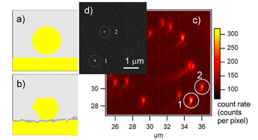
Figure 1: a) Idealized sphere-on-plane geometry b) sketch of a more realistic geometry including surface roughness, faceted crystallites and imperfect spacer layers, c) Scattering from individual sphere-on-plane structures obtained in a confocal microscope, d) electron micrograph of two selected structures as indicated.
We present experimental investigations on sphere on plane structures formed by colloidal metal particles, separated from a metal surface by an organic spacer layer. The substrate material and morphology, the spacer layer and the colloidal metal particles were systematically varied. Surface plasmon spectroscopy with multicolor excitation is used as a highly sensitive method for ensemble characterization of the resulting resonances. Optical single object investigations coupled with electron microscopy complement these studies, giving access to the homogenous line width and therefore the quality factor of the resonator.
Laboratory of Physical Chemistry, ETH Zurich, CH-8093 Zurich, Switzerland
The strength of the interaction between an emitter and radiation can be modified in the near field of a nanostructure. In particular, metallic nanostructures can be quite effective in this respect because they can support plasmon resonances, increasing their scattering cross sections. The nanostructure can act as a resonant antenna to enhance the excitation rate and the spontaneous emission rate of the emitter. However, the vicinity to the metal can also lead to an increased nonradiative decay rate of the excited state. We present experimental results on the strong modification of more than twenty times in the fluorescence intensity and decay rate of a single molecule in the near field of a single gold nanoparticle [1]. By using scanning probe techniques, the distance between the nanoparticle and the molecule is controlled while the fluorescence lifetime and intensity are measured at each pixel. In addition, experiments are presented where extended metallic or glass tips are placed in close vicinity of single molecules [2]. We also show that the interaction of a dye molecule with a gold nanoparticle can be exploited for the realization of a nanoscopic ruler with an interaction length beyond that of conventional FRET [3]. Furthermore, we discuss theoretical calculations that address the issue of losses in metals and predict a substantial improvement of these results by optimizing the geometry [4]. Finally, we put these efforts in the context of our recent measurements where the coherent interaction of a laser beam with a single molecule has been studied in the near field [5, 6].
[1] S. Kühn, U. Hakĺnson, L. Rogobete, and V. Sandoghdar, Phys. Rev. Lett. 97, 017402 (2006); see also the Supplementary Materials.
[2] S. Kühn and V. Sandoghdar, Appl. Phys. B 84, 211(2006).
[3] J. Seelig, K. Leslie, A. Renn, S. Kühn, V. Jacobsen, M. van de Corput, C. Wyman, V. Sandoghdar, Nano. Lett. 7, 685 (2007).
[4] L. Rogobete, F. Kaminski, M. Agio, V. Sandoghdar, to appear in Opt. Lett.
[5] I. Gerhardt, G. Wrigge, P. Bushev, G. Zumofen, M. Agio, R. Pfab, V. Sandoghdar,
Phys. Rev. Lett. 98, 033601 (2007).
[6] I. Gerhardt, G. Wrigge, M. Agio, P. Bushev. G. Zumofen, V. Sandoghdar, to appear in Opt. Lett.
Junior Professor for Nano-bio-technology and Emmy Noether Research Group Leader Institute for Physical Chemistry, University of Mainz, Jakob-Welderweg 11, 55128 Mainz, Germany email: soennichsen@uni-mainz.de (http://www.nano-bio-tech.de)
We present a continuous flow synthesis of gold and silver nanorods allowing the tailored design of particles with desired shape and the on-line monitoring of particle growth [1]. A novel bio-functionalization procedure for such particles is developed [2] and such particles investigated using an innovative fast single particle spectroscopy (fastSPS) method[3].
Continuous flow microfluidic reactors offer some advantages over conventional batch based methods: straight-forward up-scaling, on-line monitoring of the products for easy process control, improved reaction yield, selectivity, and kinetics by exploring a wider parameter space for external parameters like pressure and temperature, and improved reactant mixing. Miniaturization of the flow channels allows the change of experimental conditions along the reaction path within microseconds and greatly reduces the volume required to test certain experimental parameters (to microliters or even nanoliters).We show here the first example of a continuous flow synthesis of nanoparticles with a rod-like shape, specifically gold and silver nanorods.
The plasmon in nanorods splits into two modes for electron oscillations along the long and short particle axis. The optical spectrum is dominated by the much stronger long axis oscillation at longer wavelengths. The wavelength λp of this long axis plasmon depends strongly on the aspect ratio a/b of the gold rods. An empirical relation (λp/nm=(53.71a/b – 42.29)εm+495) has been derived for gold nanorods, which is useful to derive the approximate aspect ratio directly from an optical extinction measurement.
The results of a continuous flow synthesis run with varying flow rates are shown. For different mixing ratios, the gold nanorods have different aspect ratios as judged from the extinction maximum. Less seeds produce higher aspect ratio rods. Since the growth solution is depleted slower for less seeds growing into nanorods, this result should be expected. We observe a slow overall drift towards longer wavelength over the experimental time (visible in most experiments), most likely due to a slow precipitation or disintegration of the seeds in the syringes. We also test the influence of growth temperature on particle shape. Here we observe a shift of the extinction maximum towards lower wavelength for higher growth temperatures, indicating smaller aspect ratio (‘fatter’) rods. The reason for this could be an increased surface mobility of the adsorbed gold atoms or a higher permeability of the CTAB micelle for gold ions at higher temperatures.
In addition to their striking colors, noble metal nanorods are interesting materials for a range of applications including use as contrast agents in medical applications, in ultrasensitive biosensors[4], and for the enhancement of nonlinear optical effects. Rod-shaped nanoparticles often have superior optical properties compared to their spherical counterparts due to the optical antenna effect [5].
[1] Boleininger, J.; Kurz, A.; Reuss, V.; Sönnichsen, C. Phys. Chem. Chem. Phys. 2006, 8, 3824.
[2] Pierrat, S.; Becker, J.; Zins, I.; Sönnichsen, C. Nano Letters 2006, ASAP Dec 22.
[3] Becker, J.; Schubert, O.; Pierrat, S.; Sönnichsen, C. submitted 2007.
[4] Raschke, G.; Kowarik, S.; Franzl, T.; Sönnichsen, C.; Klar, T. A.; Feldmann, J.; Nichtl, A.; Kurzinger, K. Nano Letters 2003, 3, 935-938.
[5] Sönnichsen, C.; Franzl, T.; Wilk, T.; von Plessen, G.; Feldmann, J.; Wilson, O.; Mulvaney, P. Phys. Rev. Lett. 2002, 88, 077402.
International Centre of Biodynamics, Bucharest, Romania www.biodyn.ro
A novel Dual Surface Plasmon Resonance (SPR) - Differential Electrical Impedance (DEI) measurement set-up is used to investigate the capabilities of differential impedance spectroscopy to assess bioaffinity sensors (with the surface modified with high specificity compounds) for both direct and competitive determinations, aiming for development of an effective assay for target analytes detection. The surface changes due to the specific recognition events are simultaneously monitored, on the surface of the same chip using this custom designed system that comprises: an SPR module, a multi-channel, differential impedance analyzer, a fully automated injection system (with capabilities for manual injecting and preprogrammed protocols) and a 2 channel micro-fluidic, dual measurement chamber.
In this format, the variations of sensor/sample interface in response to fast specific recognition events (Ag-Ab) are revealed, while eliminating the nonspecific influences by simultaneous SPR and DEI measurements providing inner validation and expanding the analyte detection range.
The system was successfully tested for concentration analyses of a wide range of target analytes from low molecular analytes (e.g. gentamicin) to whole cells.
Institute for Photonic Technology Jena
The plasmonic properties of nanoscale metal structures depend on parameters such as material composition, dimensions, shape and optical properties of the surrounding media. Therefore designer particles with a chosen absorption band are possible by e.g. choice of the material composition in the case of core-shell particles. We present here work towards the synthesis of core-shell particles using Au/Ag and Ag/Au systems. The optical properties were determined both on an ensemble level using UV-VIS spectroscopy [1] as well as in a single particle approach in order to determine the scattering spectra of individual particles. This single particle approach can be combined with AFM imaging of the same particle even inbetween several steps of e.g. shell growth. The change of the surrounding media induces shifts in the absorption band position that can be used in sensoric applications.
[1] A. Steinbrück, A. Csaki, G. Festag and W. Fritzsche, Plasmonics 1 (2006) 79.
1Institute of Physical Chemistry, Friedrich-Schiller-University Jena, Helmholtzweg 4, D-07743 Jena, Germany
2Institute of Photonic Technology, Albert-Einstein-Str. 9, D-07745 Jena, Germany Jürgen Popp, email: Juergen.Popp@uni-jena.de
In medical applications e. g. drug delivery there is the demand to determine the concentration of analyts (for example antibiotics or narcotics) online with a small sample volume and without any external markers. Applying micro-Raman spectroscopy a spatial resolution of approximately 1 µm can be achieved. One disadvantage of Raman spectroscopy is the low sensitivity compared to other techniques.
Applying Surface Enhanced Raman Spectroscopy (SERS) the sensitivity can be highly increased.[1] However a quantitative assessment is difficult as the conditions like mixing and activation time have to be kept constant. In combination with micro fluidic devices, where all these parameters are kept constant, fluctuations in the concentration of special analytes can be quantitatively monitored online. This is very interesting when investigating the concentration of water pollutants in drinking water or the drug concentration in blood serum.[2, 3]
In the following contribution we present the SERS detection of aqueous analytes like drugs in water droplets embedded in a stream of oil. Via injection ports the aqueous analyte solution as well as the colloidal solution is directed into a stream of tetradecane. The advantage of this so called segmented flow is that the adhesion of nanoparticle aggregates to the channel walls is prevented by a thin film of oil which surrounds the water droplets.[4]
The flow velocity of the oil and the injected aqueous droplets can be regulated with an external syringe pump system. For the detection the laser of a micro Raman setup is focused directly into the channel and the signal is detected in backscattering geometry.
Acknowledgement
Financial support of the Deutsche Forschungsgemeinschaft (DFG; grant number Po 563/4-1) and the BMBF/VDI (16SV1998, SERIZELL) is gratefully acknowledged.
[1] Nie, S.; Emory, S. R., Probing single molecules and single nanoparticles by surface-enhanced Raman scattering. Science (Washington, D. C.) 1997, 275, (5303), 1102-1106.
[2] Lee, D.; Lee, S.; Seong, G. H.; Choo, J.; Lee, E. K.; Gweon, D.-G.; Lee, S., Quantitative analysis of methyl parathion pesticide in a polydimethylsiloxane microfluidic channel using confocal surface-enhanced Raman spectroscopy. Applied Spectroscopy 2006, 60, (4), 373-377.
[3] McLaughlin, C.; MacMillan, D.; McCardle, C.; Smith, W. E., Quantitative Analysis of Mitoxantrone by Surface-Enhanced Resonance Raman Scattering. Analytical Chemistry 2002, 74, (13), 3160-3167.
[4] Strehle, K. R.; Cialla, D.; Rösch, P.; Henkel, T.; Köhler, M.; Popp, J., A Reproducible Surface-Enhanced Raman Spectroscopy Approach. Online SERS Measurements in a Segmented Microfluidic System . Analytical Chemistry 2007, 79, (4), 1542-1547.
1IPHT Jena, Albert-Einstein-Straße 9, D-07745 Jena, Germany
2JBCI, IPC / Friedrich-Schiller-University Jena, Helmholtzweg 4, D-07743 Jena, Germany
Surface enhanced Raman scattering (SERS) is an ultra sensitive analytic tool for the detection of low concentrated biological or chemical analytes using the enhancement of electrical fields at metallic nanostructures. For easy sample handling as well as for quantization, e.g. the determination of the analyte concentration, arrays of sharp-edged metallic structures < 100 nm with stringent control on the enhancing feature shapes are indispensable. The preferable array size for automized measurement is about 200 × 200 µm2.
We have fabricated arrays of star-like and diamond-like gold patterns placed on quartz substrate using electron beam lithography and ion beam etching. The star-like gold pattern (thickness 25 nm) with a period of 250 nm (corresponding to 800 × 800 stars) showed smallest structure details of about 20 nm in width. The micro-fabricated SERS-substrates were tested using crystal violet as test substance measuring SERS and Raman spectra. From first measurements the calculated enhancement is about 102 using the star-like.
1Graduate Institute of Electro-Optical Engineering, National Taiwan University, Taipei, Taiwan 10617
2Center of Nanostorage Research, National Taiwan University, Taipei 10617 Taiwan
3Department of Physics, National Taiwan University, Taipei 10617, Taiwan
4Research Center for Applied Sciences, Academia Sinica, Taipei 115, Taiwan
Nanoscale metallic particles possess the fantastic characteristics of interacting with light to induce multipolar surface plasmon resonance (SPR) modes under proper conditions. By varying the materials, the geometrical shapes, and the surrounding medium of metallic nano-particles, the resonant wavelength of the SPRs can be efficiently tuned leading to successful applications of nano-antennas, bio-sensors, and nano-imaging. In addition, the enhancement of light by metallic nano-particles also contributes to the development of SERS and near-field optical disk storage. Since the metallic nano-particles can function as fundamental building blocks in plasmonic nanophotonics, it is crucial to know their individual optical properties.
In this paper, the growth of gold nanoparticles by CTAB template is performed in a finite volume solution on the top of a glass slip. A polarization-contrast microscopy coupled with an atomic force microscope is utilized to attain far-field optical images of the multipolar SPR modes of a single gold nano-particle. The topographic measurements and optical observations of the samples can be studied simultaneously. During the growth process of metallic nano-particles, the dynamic optical responses clearly indicate the variations of the plasmonic modes induced by the incident light. We also found a liner relationship between the wave vectors of the parallel incident light and the induced SPR modes on a single gold nano-rod. Besides, we can arrange a metallic nano-particle chain by the AFM and obtain its plasmonic optical properties with the same instrument. Our experimental results demonstrate a feasible way on acquiring plasmonic optical properties from an individual single gold nano-particle and assist the development of novel applications of plasmonic nanophotonics.
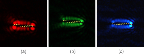
Far-field images of SPR modes of the same single gold nano-rod for various incident laser light at (a) red (658 nm), (b) green (532 nm), and (c) blue (488 nm), respectively.
Philipps University of Marburg, Dept of Physics, Applied Physics Renthof 7, D-35032 Marburg, Germany
Inorganic hydrophobic nanoparticles of different materials such as Au, CdSe/ZnS, CoPt etc. can be coated with an amphiphilic polymer to yield particles that are stable in aqueous solution.The carboxylic groups on the surface of the polymer shell serve as anchor points for further chemical functionaliziation. Ligand molecules with amino groups can be covalently bound to the particles. Poly(ethylene glycol) (PEG) is an inert biocompatible polymer that is known to decrease unspecific binding of particles to surfaces and to increase the collidal stability at physiological salt concentrations. With bifunctional PEG molecules, the particles can be modified with additional functional groups such as amines, thiols, maleimides etc.By the increase in size, the binding of the PEG molecules to the particles can be monitored by gel electrophoresis and other techniques. If the molecular weight of the PEG molecule is high enough, conjugates of nanoparticles with one, two, and three PEG molecules per nanoparticle can be separated using gel electrophoresis. In this way the PEG molecules act as spacers that allow the sorting of nanoparticles with a discrete number of functional groups, in order to eliminate uncontrolled inter-particle crosslinking in further experiments.