Home | Photos | Focus | Program | Scientific committee | Abstracts | Travel | Registration | Past Meetings

Download abstracts as PDF-file.


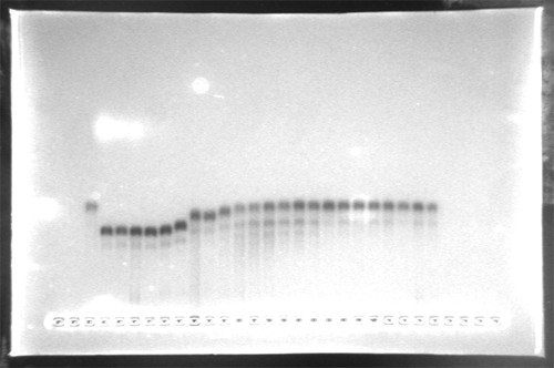
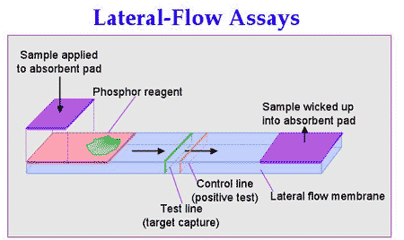
Oligonucleotide Conjugated Gold Particles for Genomic Analysis

Y. Paul Bao, Sudhakar S. Marla, James J. Storhoff, Tai-Fen Wei, Susan Hagenow, Hitesh Mehta, Adam Lucas, Viswanadham Garimella, William Cork and Uwe Müller

Nanosphere, Inc., Northbrook, IL 60062, USA

Fluorescent labels, either incorporated directly into the target sequence or attached indirectly, represent the basis for the currently preferred labeling scheme in biomolecule detection and array applications, but an increasing emphasis on higher sensitivity has led to the development of dendrimers, quantum dots, up-converting phosphors and nanoparticles.Silver amplified 13 nm gold-nanoparticle probes have been shown to allow high sensitivity detection since they can be detected by light scatter at a density as low as ~0.0025 probes/um2, using Nanosphere's extremely low-cost evanescence-based image analysis system. This is on the order of 1000-fold more sensitive than laser scanning-based fluorescence detection on a particle to molecule basis. We have developed a highly effective method for functionalizing nanoparticles with modified oligonucleotides. The oligo-coated nanoparticles result in probes of high chemical and thermal stability, low non-specific binding, and sharp melting transitions. These probes can hybridize to nucleic acid targets under conditions of significantly elevated stringency and achieve high specificity. We have applied this remarkably specific and sensitive nanoparticle-based analysis system to the development of microarray based research as well as diagnostic assay systems. Preliminary data suggest attomolar sensitivity when testing for infectious agents and genetic predispositions in model systems with single-nucleotide discrimination. In fact, our probes show sufficient discrimination power to differentiate between unique single nucleotide polymorphisms in the presence of total human DNA. Furthermore, we have successfully developed a universal nanoparticle probe system that allows label-free mRNA detection in expression arrays and have observed more than 40-fold increased sensitivity.


Fluorescent and magnetic nanoparticles for molecular diagnostics of nucleic acids on a single molecule level

Frank F. Bier, Ralph Hölzel, Nenad Gajovic-Eichelmann, Alexander Christmann

Fraunhofer Institute for Biomedical Engineering, Dept. Molecular Bioanalytics, Bergholz-Rehbrücke, Germany

Analytic and clinical diagnostic on a single molecule level is of great interest in all that cases where only few or even single cells are to be investigated. Especially on the genomic level e.g. for the analysis of transcriptomes single events are ruling the behaviour of the cell as a whole and might be crucial for the formation of cancer. Nanoparticles are an alternative method to enhance signals linked to single molecular binding events. In our studies we use stretched long DNA strands of several µm length (< 7000 bp). As has been reported previously long doublestranded DNA may be stretched by virtue of alternating electric fields. Recently we found that also single stranded DNA may be stretched in the same way. Defined sites on stretched DNA can easily be addressed by an oligonucleotide that is complementary to this site. To visualise such single oligonucleotides fluorescent particles enhance the signal compared to fluorochrome label by several orders of magnitude. An alternate label are magnetic particles of 50 nm diameter. This type of particles are well distinguishable by Magnetic Force Microscopy (MFM) with an extremely high signal to noise ratio. Moreover, such labels are also used as construction tools to direct the captured end of a long DNA fragment to a defined position.


Inorganic Nanoparticles in Medical Diagnostics

K. Bohmann*, W. Hoheisel*, S. Haubold#, C. Meyer#, M. Haase+,

*Bayer AG, D-51368 Leverkusen, Germany; #Nanosolutions GmbH, D-22525 Hamburg, Germany; +Phys.-Chem. Institut, Univ. Hamburg, D-20146 Hamburg, Germany

Advances in producing and handling nanoscaled materials lead to increasing commercial interests in the use of these materials. One of the first sophisticated nano-products could be introduced in medical diagnostics for labeling biomolecules. Immunoassays, biosensors or PCR processes often rely on fluorescent labels to quantify analytes like antibodies or DNA-strands. Organic molecular dyes still dominate this field even though they show some crucial drawbacks like fading or limited capabilities to detect several analytes in parallel (multiplexing). Due to the need for alternative approaches a number of new technologies is being developed employing gold colloids or quantum dots. The use of nano-sized, phosphorescent particles is a less known approach for fluorescent biolabels even though they show decisive advantages. Nanophosphors are environmentally acceptable nanocrystals like LaPO4, Y2O3 or sulfates being doped with Rare Earth ions. Upon UV-excitation they fluoresce with a characteristic, narrow banded wavelength pattern which is independent of the particle size. This again strongly facilitates large scale production. Furthermore, long living excited states with half-lifes in the millisecond range allow time resolved measurements with increased sensitivity due to background free detection. It could be demonstrated that nanophosphors can be quenched via fluorescence resonance energy transfer (FRET) to attached dyes. Exemplified by oligonucleotides coupled to LaPO4:Ce,Tb we could demonstrate the feasibility of homogeneous assays which renders nanophosphors superior over other particle based systems. For any new biolabel system this is an important condition for a broad use outside market niches.


Optical detection of individual nanoparticle labels after metal enhancement

Andrea Csáki,*, Pia Kaplanek, Wolfgang Fritzsche

Biotechnical Microsystems Department, Institute for Physical High Technology, Jena, Germany

The detection of DNA using nanoparticle label is an interesting alternative to the standard fluorescence technique. It requires simpler detection equipment, resulting in higher stability and lower costs. Beside easier detection, metal enhancement results in a higher sensitivity of detection. The signal-response curve for labeled DNA before and after silver enhancement was studied, applying both AFM and optical (reflection/absorption) measurements. So the dynamic range and the sensitivity were determined for nanoparticle labeling with and without metal enhancement. The ultimate limit of detection, the identification of individual labels, is demonstrated for the optical readout of metal-enhanced gold nanoparticle. Therefore, chosen nanoparticles were visualized before and after enhancement, so that the individual optical signals could be clearly related to either single or multiple nanoparticles.


Gold nanoparticle labels in electrochemical ligand-binding assays:application to the detection of viral DNA hybridization

Murielle Dequaire, Céline Grossiord, Laurent Authier, Benoít Limoges, Pierre Brossier

Laboratoire de Microbiologie Médicale et Moléculaire, Facultés de Médecine et de Pharmacie, 7 Boulevard Jeanne d'Arc, 21000 Dijon, France

Gold nanoparticles labels have gained considerable interests in the development of ligand-binding assays (immunoassays, hybridization tests) because many analytical methods are suitable for their quantitative detection, e.g., colorimetric absorption spectrophotometry, microgravimetry, scanning force microscopy, surface plasmon resonance...

Recently, we have developed a novel electrochemical amplification strategy for the sensitive detection of colloidal gold nanoparticles by determining the amount of AuIII ions released in solution from each nanoparticle after oxidative treatment in an acidic solution [1]. For this purpose, we have employed the highly sensitive anodic stripping voltammetry (ASV) at a disposable carbon-based screen-printed electrode. An increase of sensitivity was obtained at a screen-printed microband electrode (AuIII detection limit of 5 x 10⁻⁹ M) which allows for the enhancement of AuIII ions mass transfert and thus for simplified measurements in small volumes of quiescent solution [2].

Here the sensitive Au nanoparticle-based electrochemical detection is applied to the determination of an amplified 406-base pair human cytomegalovirus DNA sequence (HCMV DNA). The principle of the assay, which is an adaptation of the commercially available Hybridowell® test (http://www.argenebiosoft.com), consists of four main steps sketched below: (a) passive adsorption of the HCMV-amplified DNA target on the wall of polystyrene microwell, (b) hybridization with a 22-base oligonucleotide probe conjugated to a 20-nm colloidal nanoparticle, (c) oxidative gold metal dissolution in an acidic bromine-bromide solution, and (d) ASV detection of the released AuIII ions at a SPMBE immersed in the microwell.

The analytical performance of this gold nanoparticle-based electrochemical HCMV-DNA hybridization assay will be discussed and compared with previously reported HCMV DNA hybridization tests involving an enzyme label [3].

1. Dequaire M, Degrand C, Limoges B. Anal Chem 2000; 72, 5521-8.
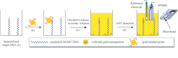
2. Authier L, Grossiord C, Brossier P, Limoges B. Anal Chem 2001; 73, 4450-6.
3. Azek F, Grossiord C, Joannes M, Limoges B, Brossier P. Anal Biochem 2000; 284, 107-13


Substrate modification for gold nanoparticle labeling

Grit Festag, Andrea Steinbrück, Andreas Wolff, Andrea Csáki, Robert Mölller, Wolfgang Fritzsche

Biotechnical Microsystems Department, Institute for Physical High Technology, Jena, Germany,

Immobilized DNA can be detected by complementary, nanoparticle-labeled DNA. For parallel readout, optical and electrical detection methods have been introduced. Silver enhancement of gold nanoparticles allows the optical readout by standard flatbed scanners. However, substrate modification, enabling DNA binding, could lead to inhomogeneous surfaces and unspecific bindings of the gold particles. Contact angle measurements as well as fluorescence, AFM and a flatbed scanner were used to analyze the multiple step process, including substrate surface modification, immobilization of the capture DNA, specific binding of the gold-labeled DNA probe, and their enhancement by silver deposition.


Labeling Biomolecules with Nano Metal Particles

James F. Hainfeld

Nanoprobes, Inc., 95 Horseblock Rd., Yaphank, New York, USA 11980; & Brookhaven National Laboratory, Bldg. 463, Upton, New York, USA 11973

The early history of labeling biomolecules with metal clusters will be presented, as well as recent developments. Labeling of various proteins, peptides, nucleic acids, lipids, and other molecules using small, well-defined gold compounds is possible with specific coupling chemistry. The types of metal clusters can be varied, and include gold, platinum, silver, iridium, tungsten, and other metals that form nanoparticles.

Another related technology is autometallography, where the initial metal nanoparticle serves as a nucleation center for additional metal deposition, typically of silver, but may also include gold, copper, and other metals. This grows the small particles into more detectable sizes, thus increasing sensitivity.

Gold labeled proteins may also be combined with fluorophores to produce dual probes useful for correlative light and electron microscopy.

There are many uses of these technologies, such as immunohistochemistry, in situ hybridization (even detection of single gene copies), nanomaterials, nanoelectronics, disease detection, bioterrorism detection, cell and molecular studies, gene transfection, gene and enzyme control, and in vivo applications, just to name a few.

DNA Nanowires: 1.4 nm Nanogold clusters (bright spots) bound to double stranded bacteriophage T7 DNA (rope-like strands). Dark field, unstained STEM image on a thin carbon substrate. Full width 128 nm.


Magnetic nanoparticles for the treatment of tumours by magnetic heating

Ingrid Hilger

Institute for Diagnostic and Interventional Radiology, University Hospital Jena, Germany

The therapeutic strategy for the treatment of tumours is being changing in the last time. Particularly for breast cancers, an increasing incidence of small tumours with good prognosis is being detected due to the improvements of diagnostic methods. Radical mastectomy (the total excision of the breast) is being applied only in cases with multicentricity, multifocality, tumour spreading over several quadrants or extensive infiltration. In contrast to that, breast conserving therapies are gaining an increased significance. Newer investigations are dealing with further steps towards the minimal-invasive elimination of tumours.
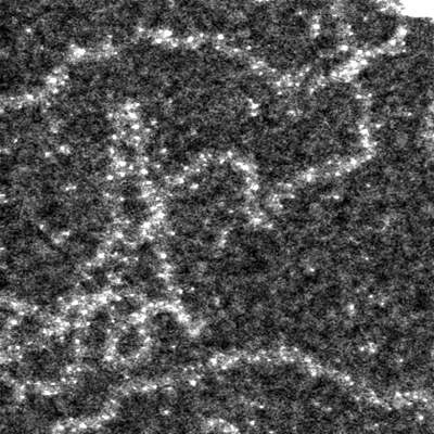
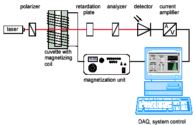
Considering this aspects, the accumulation of a magnetic material (iron oxides, e.g. magnetite) in the tumour region and the exposure of the whole breast to an alternating magnetic field was proposed. By this procedure, the magnetic material absorbs energy from the magnetic field and converts it into heat which is used to eliminate the tumour.
The elaboration of the proposed therapy conduced in a close co-operation with the Institute of Physical High Technology (Jena, Germany) method will be presented. We describe the heating potential of magnetic materials, the particle interaction with cells, the choice of magnetic field parameters taking into account the effect of unfavourable eddy current heating. We further report the functional dependencies for the expected temperature increase at the tumour site, the critical heat dose for a destruction of tumour cells, in vivo- and in vitro experimental data and numerical estimations for the feasibility of the generation of localised heat spots. The expected wash out of magnetic material from the tumour region is discussed on the base of our experimental findings.


Conjugation of Au-nanoparticles with DNA

Almudena Muñoz Javier, Teresa Pellegrino, Stefan Kudera, Wolfgang J. Parak

Center of Nanoscience, Ludwig-Maximilians-Universität München, Amalienstra§e 54, Germany

For optical investigations of the quenching of fluorophores on gold surfaces it is important to control the distance between the fluorophore and the gold. For this purpose we use DNA of different length as spacer between fluorophore and gold. To do so we incubate gold nanoparticles with DNA which is functionalized at one side with the fluorophore and at the other side with a thiol-group that can bind to gold. To control the conformation of the DNA we make a series of different DNA to gold ratios and run them on an agarose gel. This is an example of this gel:

Since DNA and gold are negatively charged, the conjugates migrate towards the plus pole. The more DNA is attached the bigger the DNA-gold conjugate becomes which implies a reduced mobility. At one point even if more DNA is added the mobility is not reduced further. This means that the gold is saturated with DNA. At this point the DNA is fully stretched, and the distance between the fluorophore and the gold is equal to the length of the DNA.

Enzymatic manipulation of DNA/Gold Nanostructures

Antonios G. Kanaras*#, Zhenxin Wang*, Andrew D. Bates#, Richard Cosstick* and Mathias Brust*

Centre for Nanoscale Science: * Department of Chemistry, The University of Liverpool, Liverpool L69 7ZD, UK.# School of Biological Sciences, The University of Liverpool, L69 7ZB, UK

DNA manipulation enzymes (restriction endonucleases, ligases) have been employed to control the assembly of gold/DNA hybrid nanostructures. Using these well-established bio-molecular tools it is now possible to carry out multi-step syntheses of nanostructures following concepts, which are very similar to those known from preparative chemistry. The elemental building blocks of these structures are gold nanoparticles, which carry a number of double stranded DNA molecules in their ligand shell. The base sequences of these molecules determine whether they can be converted into reactive sites by cleavage with specific restriction enzymes. The ends of such cleaved strands are phosphate activated and can participate in a further enzyme dependent reaction, which links them covalently to other reactive sites present in the system. This second step is catalysed by a DNA ligase. It is shown that nanostructures can be assembled in multi-step syntheses by successively addressing different DNA molecules with different restriction enzymes. Reactive sites are only created when the recognition sequence required by the particular enzyme employed is present. All other DNA molecules not containing this recognition site will remain un-reactive and can be activated in further steps by enzymes that recognise different sites. This concept represents a nano-scale analogue of the use of protecting groups in preparative chemistry. It is further demonstrated that existing gold/DNA nanostructures can be dissociated by cleavage at predetermined sites using restriction enzymes.

The principles of enzyme controlled nanostructure manipulation have been established using freely aggregating systems of gold particles and DNA in solution. The same reactions can also be carried out on surface immobilised structures, i.e. covalently attached particles can be enzymatically cleaved from a surface, or particles in solution can be ligated to reactive DNA double strands bound to a surface. This can be monitored by AFM. It is believed that the introduction of these powerful tools will bring nanostructure self-assembly a bit closer to rational chemical synthesis and thus will enable the step-by-step preparation of a number of unique, new nanostructures.

Antonios G. Kanaras, Zhenxin Wang, Andrew D. Bates, Richard Cosstick, and Mathias Brust, Angewante Chemie Int. Ed., 42 (2), 191-194, 2003Towards multi-step nanostructure synthesis: programmed enzymatic self-assembly of DNA/gold systems


Biomolecule-nanoparticle conjugates for bioanalysis: Nanospheres and barcoded nanowires

Christine D. Keating

Department of Chemistry, Pennsylvania State University

Metal nanoparticles can be used as amplification or identification tags for biomolecular recognition events. In both cases, the attachment chemistry and surface coverage of biomolecules on the particle surface is critically important for retention of bioactivity. We have attached DNA and proteins to metal nanoparticles ranging from 12-nm diameter colloidal Au spheres to 6 micron long, 320 nm wide striped metal nanowires. Au nanosphere:biomolecule conjugates are useful as amplification tags, to increase sensitivity in a variety of assay formats. Metal nanowires can be encoded with submicrometer stripes by sequential electrochemical deposition within the pores of alumina membranes. The metal striping pattern, or 'barcode', is identified via the differential reflectivity of adjacent stripes using conventional light microscopy. Reflectivity-based readout of particle patterns does not interfere with the use of fluorescence for detection of analytes bound to particles by affinity capture.


Spectroscopical measurements with quantum dots

Stefan Kudera*, Tim Liedl*, Wolfgang J. Parak*, Tobias Schrader#, Markus Braun#

*Lehrstuhl für Angewandte Physik, #Lehrstuhl für BioMolekulare Optik; Ludwig-Maximilians-Universität München,

In collaboration with a spectroscopy group we attempt to gain knowledge on both the processes that occur when a quantum dot is excited and on the chemical surrounding of the crystal. The samples are investigated with different spectroscopic techniques: Standard cw-UV-Vis absorption, standard cw fluorescence spectroscopy, short time spectroscopy in the IR-range and in the visible spectrum, and continuous IR absorption spectroscopy.

By means of short time absorption (both IR and visible) spectroscopy one can observe the decay channels of the excited nanocrystals. This could present a possibility to gain knowledge on the effects that lead to the bleaching of the crystals and on the interactions of the crystals with the surrounding. Ideally one would expect an exponential decay of the excitation which is the situation of an ideal two state system. Any difference from this decay mechanism has its origin in nonradiative processes.

IR absorption spectroscopy (continuous wave) is well suited for the examination of the quantum dots' surface. After the synthesis of the particles, their surface is covered with molecules of the solvent in which they were grown, which makes the particles hydrophobic. Therefore, for making the particles water-soluble, one has to exchange the hydrophobic surfactant molecules by water-soluble molecules. With IR-spectroscopy we want to determine the number of surfactant molecules per nanocrystal and thus the efficiency of the exchange. This technique could help to improve the capping of the nanocrystals.


Silanisation of CdSe/ZnS Nanocrystals

Tim Liedl, Stefan Kudera, Wolfgang J. Parak

Lehrstuhl für Angewandte Physik, Ludwig-Maximilians-Universität München, Amalienstraße 54, Germany

We intend to use semiconductor-nanocrystals in biological applications. For example biomolecules can be labelled with these crystals and then tracked on their pathway through cells. Semiconductor-nanocrystals promise to improve this tracking compared to standard organic fluorophores as they do not bleach as fast as organic fluorescent dyes.

The CdSe/ZnS nanocrystals synthesized in our group are not water-soluble. For the envisaged biological applications it is necessary to transfer them into water. We here present our method to perform this task.

There are techniques to synthesize semiconductor-nanocrystals directly in water. But the so prepared quantum dots do not show sufficiently good stability in both their lifetime and their fluorescence for many biological applications. The main advantage of the synthesis employed in our group is that it is performed at high temperature and therefore guarantees very good lattices as defects in the lattices are healed.

The idea of the silanisation is to make the particles hydrophilic by growing a silica shell around them. The process of growing the silica-shell can be broken up into three steps.


Electrical readout for DNA hybridization assays based on nanoparticle labeling

Robert Möller, Matthias Urban, Wolfgang Fritzsche

Biotechnical Microsystems Department, Institute for Physical High Technology, Jena, Germany

Immobilized DNA can be detected by complementary, nanoparticle-labeled DNA. A novel readout scheme applying an electrical measurement of changes in the electrical resistance due to nanoparticle-induced metal deposition has been introduced (1,2). This detection scheme offers the potential for a simple (low-cost as well as robust) and highly miniaturizable method, which could be well-suited for point-of-care applications in the context of lab-on-a-chip technologies. The demonstrated apparatus allows a parallel readout of an entire array of microstructured measurement sites (3). The readout is combined with data-processing by an embedded PC, resulting in an autonomous instrument that measures and presents the results. The design and realization of such a system is described, and first measurements are presented.

[1] Möller, R., Csáki, A., Köhler, J. M. & Fritzsche, W. Electrical classification of the concentration of bioconjugated metal colloids after surface adsorption and silver enhancement. Langmuir 17, 5426-5430 (2001).
[2] Park, S. J., Taton, T. A. & Mirkin, C. A. Array-based electrical detection of DNA with nanoparticle probes. Science 295, 1503-6 (2002).
[3] Urban, M., Möller, R. & Fritzsche, W. A paralleled readout system for an electrical DNA-hybridization assay based on a microstructured electrode array. Review of Scientific Instruments 74, 1077-1081 (2003).


Magnetic nanoparticles by glass crystallisation

Robert Müller, H. Steinmetz, R. Hiergeist , W. Gawalek

Magnetics Department, Institute for Physical High Technology, Jena, Germany

Nanometer-scale particles reveal unique magnetic properties. In the last time magnetic nanoparticles became interesting for biomedical applications like cancer treatment (hyperthermia), magnetic markers and detection of biological bond reactions, respectively. In all cases the particles have to fulfil certain magnetic properties for an optimised application. Commercial magnetic beads are limited concerning some properties (range of Néel relaxation time, magnetic losses, size). We are able to prepare tailored single crystalline magnetic particles in a wide range of particle size, hysteresis parameters, relaxation behaviour and magnetic losses by means of the glass crystallisation method. The particles grow during a temperature treatment isolated in a glass matrix and were leached out by dissolving the matrix. The mean particle size in the range of 10 to 30nm can be easily influenced by the temperature treatment (in contrast to the size of magnetite by usual precipitation). Our work will show results on nanocrystalline barium hexaferrite BaFe12-2xCoxTixO19 and first results on magnetic iron oxide. Nanocrystalline Ba-ferrite powders with mean particle sizes < ca. 10 nm behave superparamagnetically and show at bigger particle sizes up to ca. 30 nm, depending on temperature and measuring frequency of the magnetic field, the transition to single domain Stoner-Wohlfarth behaviour. Beside the particle size the anisotropy energy K1.V, and thus the Néelian relaxation time, depends also on the amount x of CoTi-doping.


Site Selective 1D Arrangement of Gold Nanoparticles on DNA

M. Noyong, K. Gloddek, U. Simon

RWTH Aachen, Institute of Inorganic Chemistry, Aachen, Germany

DNA has widely been used as an assembling molecule for nanoparticles in one [1,2], two [3,4] and three dimensions [5]. Thus, it can be used as a template for the formation of metallic nanowires. Therefore, DNA has to be metallized in a controlled way, which means metal nanoparticles of defined size and spacing need to be organized along the strand. Several works demonstrated DNA metallization, but without a decisive control over the particle topology [1,6-8]. cis-Pt (cis-Pt(NH3)2Cl2) can be site selectively bound to DNA [9,10], while some binding sites are still accessible for ligand exchange.Here we report first results on a direct deposition of cysteamine stabilized 3nm Au nanoparticles on DNA strands from solution [11]. First, cis-Pt is intercalated into DNA double strands. In a second step, this is followed by a ligand exchange, whereby the amino groups at the platinum complex are replaced by the ligands of the gold nanoparticles.

Fig. 1: AFM phase image of DNA/nanoparticle assemblies on silicon. A and B show partially decorated and undecorated DNA strands, respectively.

These DNA/nanoparticle assemblies are prepared on mica and silicon and probed by AFM (Fig. 1) and TEM imaging. This shows that the one dimensional arrangements of gold nanoparticles along DNA strands can successfully be mediated by the intercalation of cis-Pt.

[1] E. Braun, Y. Eichen, U. Sivan, G. Ben-Yoseph, Nature 391, p. 775-778 (1998).
[2] A.P. Alivisatos, K.P. Johnsson, X. Peng, T.E. Wilson, C.J. Loweth, M.P. Bruchez Jr., P.G. Schultz, Nature 382, p. 609-611 (1996).
[3] S. Peschel, B. Ceyhan, C.M. Niemeyer, S. Gao, L. Chi, U. Simon, Materials Science and Engineering C 19, p. 47-50 (2002).
[4] T.A. Taton, C.A. Mirkin, R.L. Letsinger, Science 289, p.1757-1760 (2000).
[5] C.A. Mirkin, R.L. Letsinger, R.C. Mucic, J.J. Storhoff, Nature 382, p. 607-609 (1996).
[6] J. Richter, M. Mertig, W. Pompe, I. Mönch, H.K. Schackert, Appl. Phys. Lett. 78 (4), p. 536-538 (2001).
[7] W.E. Ford, O. Harnack, A. Yasuda, J.M. Wessels, Adv. Mater. 13 (23), p. 1793-1797 (2001).
[8] O. Harnack, W. E. Ford, A. Yasuda, J.M. Wessels, Nanoletters 2 (9), p. 919-923 (2002).
[9] E.R. Jamieson, S. J. Lippard, Chem. Rev. 99, p. 2476-2498 (1999).
[10] E.L.M. Lempers, J. Reedjik, Advances in Inorganic Chemistry 37, edited by A. G. Sykes (Academic Press Inc., San Diego, 1991), p. 183.
[11] M. Noyong, K. Gloddek, U. Simon, Mat. Res. Soc. Symp. Proc. 735, (2003) in press.


Biological applications of quantum dots

Wolfgang Parak

Center for Nanoscience, Ludwig Maximilians University Munich, Amalienstr. 54, 80799 Muenchen, Germany

Due to their interesting properties, research on colloidal nanocrystals (quantum dots) has moved in the last years from fundamental research to first applications in materials science and life sciences. We will discuss some recent biological applications of colloidal nanocrystals. First, the properties of colloidal nanocrystals and how they can be synthesized are described. Second, the conjugation of the nanocrystals with biological molecules is discussed. And third, three different biological applications are introduced: i) the arrangement of nanocrystal-oligonucleotide conjugates using molecular scaffolds such as single stranded DNA, ii) the use of nanocrystal-protein conjugates as fluorescent probes for cellular imaging, iii) a motility assay based on the uptake of nanocrystals by living cells.


A novel 2D invasion assay using quantum dot layers

T. Pellegrino^*, W.J. Parak*%, M. LeGros #, R. Boudreau #, D. Gerion*, C. Larabell #, G. Natile^, A.P. Alivisatos*

* Univ Calif Berkeley, Dept Chem, Berkeley, CA 94720 USA^ Dipartimento Farmaco-Chimico, University of Bari,Italy% Center for Nanoscience Ludwig Maximilians University Munich, Germany# Lawrence Berkeley Lab, Div Mat Sci, Berkeley, CA 94720 USA

The ability of tumor cells to invade surrounding tissues and to metastasize to different sites is related with the cell motility. To test invasive capacities of different cancer cell lines we have developed a two dimensional invasion assay using water-soluble CdSe/ZnS nanocrystals. Semiconductor nanycrystals are inorganic fluorophores: excitation with UV light stimulates fluorescence in the visible range, whereby the color of fluorescence can be controlled by the size of the particles. It has been shown that cells are able to engulf nanocrystals in non-specific way. When cancer cells are seeded on top of a homogenous nanocrystals layer and are incubate at 37 oC for 24 h, they create trails free of nanocrystals that are no longer fluorescent. The size and the shape of these trails are related with the potential of invasiveness of the cells. We have compared the behavior of six different human cancer cell lines including colon, breast, lung and bone cancer cells. With this test it is possible to discriminate between non-invasive and invasive cancer cell lines.


Synthesis of highly fluorescing CdSe/ZnS nanocrystals

Elias Puchner, Stefan Kudera, Tim Liedl, Wolfgang Parak

Lehrstuhl für Angewandte Physik, Ludwig-Maximilians-Universität München, Amalienstraße 54, Germany

Semiconductor nanocrystals are highly fluorescent. Compared to molecular fluorescence dyes, they stand out in the small bandwidth of their fluorescence line and in their variety of color spanning the whole visible spectrum.

Their synthesis is divided into two parts:
(1) The growth of the CdSe cores. The size of the nanocrystals determines the fluorescence wavelength.
(2) The coating of the cores with Zn-S layer to enhance quantum efficiency.

The fluorescence of the nanocrystals is caused by radiating recombinations of an electron - hole pair (exciton). Incorporation of oxygen would open channels for nonradiative recombination that would lower the intensity of fluorescence. To prevent oxidation of the crystals, we synthesize them under an argon atmosphere in an organic solvent. The solvent we use allows high temperatures (300°C) for the reaction. At that temperature the nanocrystals' size can be comfortably controlled via the reaction time that is for example around 5 minutes for green fluorescing crystals.

Despite the argon atmosphere and the pure chemicals there are still possibilities for the exciton to recombine nonradiativly, especially on the surface of the CdSe cores. To reduce this effect we grow a ZnS shell on the CdSe core that keeps the exciton away from the surface. This results in an increase of the fluorescence intensity by a factor of up to 25 that can even be seen with the naked eye. When the ZnS layer gets too thick, tension is created in the layer and there is a risk of rupture of the layer. Therefore we determine an optimal thickness of the shell for each size of the CdSe core:
After having added small amounts of stock-solution containing Zn and S to the dissolved CdSe cores, we take samples which are normalized to the same optical thickness by means of absorption spectroscopy and analyze them with a fluorescence spectroscopy.
Because the shell enlarges the space where the exciton can be located, it also lowers the recombination energy, which leads to a small redshift of the fluorescence line.

With our method it is possible to produce high-quality fluorescing ZnS coated CdSe semiconductor nancrystals.


DNA-mediated Assembly of Metal Nanoparticles: Hybridization Step and Optical Properties

Ulrike Rehn, Petra Göring, Ralf Wehrspohn, and Ulrich Gösele

Max Planck Institute of Microstructure Physics, Halle, Germany

DNA or synthetic short oligonucleotide sequences, respectively, could be used as linker molecules for metal nanoparticles. In future such a combination of technological nanostructures and molecular biology will enable new devices in medicine such as diagnostic tools or in physics to auto-assemble 3D objects. Our aim is it to create well-defined three-dimensional building blocks consisting of noble metal nanowires and colloids which are linked by DNA in a controlled way.
We have developed over the last 5 years processes that allow us to prepare highly ordered pore arrays with pore sizes in the range of 25 nm to a few microns. These pore arrays consists of either silicon or alumina. The pore arrays can be replicated to create monodisperse metal nano- or µ-wires (Ag, Au, Ni, Co, Cu) which could be used for DNA linking.
We are going to present first results on different nanoparticle-DNA combinations and optical measurements. In particular, we are going to discuss the hybridization step which is a critical process step for molecular biology and nanobiotechnology.


Nano and micro metal particles for molecular detection

José Remacle*, Isabelle Alexandre*, L.M. Hagelsieb#, Remi Pampin# and Denis Flandre#

*Laboratoire de Biochimie et Biologie Cellulaire, Facultés Universitaires Notre-Dame de la Paix; Namur, Belgium. Email: Jose.remacle@fundp.ac.be;#Research center on micro and nanoscopic materials and electronic devices, Microelectronics Laboratory, Université catholique de Louvain, Belgium.

The detection of DNA or protein in micro format is a challenge for the traditional chemical or physical methods given the constraints imposed by the fragility of the biological molecules and by the level of sensitivity required in order to make such assay useful for practical applications. We have developed a method which is based on the use of nanogold particles together with a precipitation of silver in order to obtain both the sensitivity and the specificity necessary for DNA probes as well as protein detection in microformat such as microarrays. We will show that the silver can be detected both based on its reflective or diffractive optical properties as well as on its electrical conductance, using appropriate sensors.


Magneto-optical relaxation measurements on bioconjugated nanoparticles for the determination of binding reactions

E. Romanus*, C. Groß*, G. Glöckl*, A. Reinhard#, P. Weber# and W. Weitschies*

*Ernst-Moritz-Arndt-Universität Greifswald, Institut für Pharmazie, F.-L.-Jahn-Straße 17, D-17487 Greifswald;# Friedrich-Schiller-Universität Jena, Institut für Festkörperphysik, Helmholtzweg 5, D-07743 Jena

Ferrofluids become birefringent when a magnetic field is applied perpendicular to the optical axis of light impinging the fluid, as the magnetic nanoparticles contained in the ferrofluid tend to align in the direction of the external field (Cotton-Mouton-effect). After switching off the magnetising field one can observe a relaxation of the birefringence. It has also been demonstrated that such magneto-optical relaxation measurements can be used for the determination of the binding of streptavidin with biotin by using MNP conjugated with streptavidin and biotinylated bovine serum albumin as cross-linking component [1]. Recently, we have evaluated this novel approach for the determination of binding reactions using MNP conjugated with antibodies [2]. The experiments confirmed that the determination of the relaxation of the transient field-induced birefringence of MNP can be used as a novel tool for the investigation of biological binding reactions as long as these reactions result in an increase of the particle size of magnetic nanoparticles due to aggregation.

The presented poster will focus on technical aspects of the improvement of the measurement setup.The measurement setup for the determination of the magneto-optical relaxation of ferrofluids (MORFF) consists of a laser, a polariser, aligned orthogonal to an analyser and at 45¡ to the magnetic field axis, a retardation plate with its slow axis parallel to the polariser, a cuvette containing the sample, and a detector mounted on an optical bench. The cuvette is placed into a solenoid generating a variable pulsed magnetic field of up to 10 kA/m with different magnetisation times in the range of 1ms to 500ms. After switching off the magnetising field the relaxation of the birefringence is recorded by a PIN-photodiode connected to a variable-gain low-noise current amplifier. The sytem is controlled by a PC running LabVIEW®. A high-speed (20MHz) multi-function data acquisition board records and processes the data. FigureÊ1 shows a schematic of the setup.

Fig. 1. MORFF measurement setup

The authors like to thank S. Prass, R. Bark, P. Engelhardt for technical support and helpful discussions. This research project is supported by the Deutsche Forschungs-gemeinschaft (DFG), No. WE 2555/1-3.

[1] E. Romanus, C. Groß, R. Kötitz, S. Prass, J. Lange, P. Weber, W. Weitschies, Magnetohydrodynamics, vol. 37 (2001), no. 3, pp. 328-333[2] E. Romanus, C. Groß, G. Glöckl, P. Weber, W. WeitschiesJ. Magn. Magn. Mat., vol. 252 (2002), pp. 384-386.


Magnetic nanoparticle relaxation measurement as a binding specific technique for in vivo diagnostics

E. Romanus*#, M. Hückel+, C. Groß#, S. Prass*, W. Weitschies#, R. Bräuer+ and P. Weber*

* Institute of Solid State Physics, Friedrich-Schiller-Universität Jena, Helmholtzweg 5, D-07743 Jena#Institute of Pharmacy, Ernst-Moritz-Arndt-Universität Greifswald, Jahnstraße 17, D-17487 Greifswald+ Institute of Pathology Friedrich-Schiller-Universität Jena, Ziegelmühlenweg 1, D-07743 Jena

It has been demonstrated [1], that single domain magnetic nanoparticles can be used as magnetic labels for the detection of the binding of antibodies by measuring the relaxation of their magnetization after switching off a magnetizing field, a method that is also called magnetorelaxometry (MRX). A measurement system for spatially resolved relaxation measurements of magnetic nanoparticles (MNP) was developed and tested with regard to its applicability for in vivo diagnostics. The system is based on SQUID sensors to detect the magnetic relaxation of MNP and has been optimised to operate in a laboratory environment without any magnetic shielding. Due to a dead time between switching off the magnetising field and starting the measurement only bound magnetic nanoparticles with appropriate Néel relaxation times contribute to the measured signal [2, 3]. In phantom experiments we were able to localise a phantom with a volume of volume 8 cm3 containing fixed nanoparticles with a total amount of iron of 1 µmol in 1 l of an aqueous environment containing a 1000 fold higher amount (1 mmol Fe) of the identical magnetic nanoparticles in colloidal solution. This result demonstrates the specifity of (MRX) for bound nanoparticles.In the animal experiments after injection of biologically compliant amounts of dextran coated magnetic nanoparticles a strong magnetic relaxation signal could be detected in the ventral region of the mice in vivo (Fig. 2). The post mortem investigations yielded, that the strong relaxation signals derived from the livers and spleens of the animals. This finding is in good agreement with the well known accumulation of magentic nanoparticles by the phagocytes in liver and spleen.

Fig. 1 Photograph of the anaesthetized laboratory animal with superposed relaxation signal (35 pT)

It could be shown, that spatially resolved MRX is a promising novel technique that holds the potential for imaging of the in vivo binding sites of substances labelled with magnetic nanoparticles. MRX might be suitable to overcome one of the most serious imaging problems in molecular in vivo diagnostics: the background signal caused by unbound labels still circulating in blood.

[1] W. Weitschies, R. Kötitz, T. Bunte, L. Trahms, Pharm. Pharmacol. Lett. 7 (1) (1997) 5-8.[2] J. Schambach, L. Warzemann, P. Weber, IEEE Trans. Appl. Supercond. 9 (1999), 35273530.[3] L. Warzemann, J. Schambach, P. Weber, W. Weitschies, R. Kötitz, Supercond. Sci. Technol. 12 (1999), 953-955.


- Resonant nano-cluster technology -From optical coding and high quality security features to biochips

G. Bauer+ and T. Schalkhammer*#

* Institute of Analytical Biotechnology, Technical University Delft, The Netherlands# Institute of Biochemistry and Molecular Cell Biology, University Vienna, Austria + NovemberAG, Erlangen, Germany

Metal nanoclusters exhibit high local field enhancement and nanoscale resonant behavior when exited by electromagnetic radiation. A variety of phenomena had been found, described and applied in a number of novel nanotec and bionanotec devices. Decisive for these types of devices and sensors is the precise nanometric assembly coupling the local field surrounding a cluster to allow resonance with other elements interacting with this field. In particular, the distance cluster - mirror or cluster - fluorophore gives rise to a variety of enhancement phenomena. Depending on the desired application this 'resonance' - distance is tuned from five up to 400 nm.High throughput transducers using metal cluster resonance technology are based on surface-enhancement of metal cluster light absorption (SEA). These devices can be used for detection of biorecognitive binding as well as structural changes of nucleic acids, proteins or any polymer. The optical property for the analytical application of metal cluster films is the so-called anomalous absorption. An absorbing film of clusters is positioned at 10-400 nm to an electromagnetic wave reflecting layer. At a well defined distance of the cluster to the mirror the reflected electromagnetic field has the same phase at the position of the absorbing cluster as the incident fields. This feedback mechanism strongly enhances the effective cluster absorption coefficient. The system is characterised by a narrow reflection minimum whose spectral position shifts sensitively with the interlayer thickness, because a given cluster-mirror distance and wavelength defines the optimum phase. Based on our SEA-phenomenon (licensed to and further developed and optimized by NovemberAG, Germany Erlangen) a number of commercial products had been constructed. Brandsealing® uses the patented SEA cluster technology to produce optical codings. Cluster SEA thin film systems show a characteristic color-flip effect and are extremely mechanically and thermally robust. This is the basis for its application as an unique security feature. The specific spectroscopic properties as e.g. narrow band multi-resonance of the cluster layers allow the authentication of the optical code which can be easily achieved with a mobile hand-held reader developed by november AG and Siemens AG. Thus, these features are machine-readable which makes them superior to comparable technologies.Brandsealing® is available in two formats: as a label for tamper-proof product packaging, and as "direct brandsealing® ", where label and logo are permanently applied directly and unremovable to the product surface. Together with Infineon Technologies, the SEA technology is currently developed as a direct labeling of SmartCards. For large scale production november AG and HUECK FOLIEN one of world's leaders in the field of technical films for security applications cooperate in the field of product security and brand protection. On the basis of the SEA cluster technology and the know-how of HUECK FOLIEN regarding engineered industrial films, new system solutions for security features had been developed. As a new and unique feature SEA labels had been presented at the Banknote 2003 international congress as a novel high quality security feature for banknotes.


Detection of DNA with functionalized magnetic markers

J. Schotter*, H. Brückl*, G. Reiss*, P.B. Kamp#, A. Becker#, A. Pühler#

*Department of Physics, #Department of Biology, University of Bielefeld, Germany

Compared to the established fluorescent labeling method, the use of magnetic markers in biochip sensors has important advantages with respect to the detection of biomolecules at low concentrations [1]. Additionally, the direct availability of an electronic signal allows the design of inexpensive integrated detection units. The magnetic markers are commercially available superparamagnetic microspheres. We demonstrate the applicability and functionality of Giant Magnetoresistance (GMR) sensors for detecting magnetic markers [2]. The GMR-sensors consist of patterned Cu/NiFe multilayers in the second antiferromagnetic maximum. Hybridization experiments show that the magnetoresistive biosensor can detect complex DNA with a length of about one thousand base pairs down to a concentration of 10 pg/µl. A direct comparison of the magnetoresistive and fluorescent detection methods shows that the magnetoresistive biosensor is superior to standard fluorescent detection at low concentrations.

[1] M.M. Miller et al., J. MAGN. MAGN. MATER. 225, 138 (2001)
[2] J. Schotter et al., IEEE T. MAGN. 38 (5), 3365 (2002 )


Infrared up-converting phosphors for bioassays

Hans Tanke, Michel Zuiderwijk, Paul Corstjens

Department of Molecular Cell Biology; Leiden University Medical Center, The Netherlands; (supported by OraSure Technologies, Bethlehem, PA)

Up-Converting Phosphor Technology (UPT), utilizing rare earth doped ceramic particles as biological reporters, represents the new generation of particle-based bioassays. Unlike conventional fluorescent reporters, up-converting phosphors can transfer low energy IR radiation to high energy IR or visible light by multi-photon absorption and subsequent emission of dopant-dependant phosphorescence (see Figure). Because no biological matrix possesses this unusual luminescent property, the inherent autofluorescence, confounding most fluorescence-based methods, is completely absent in up-converting phosphor assays. Enhanced assay sensitivities have been demonstrated in UPT applications, where up-converting phosphor reporters deliver 10 to 100 fold of better detection limits than conventional fluorescent probe. Up-converting phosphors can generate large anti-Stokes shifts up to 500 nm. Emission and excitation bands of these biological reporters are well separated, making the detection of phosphorescence interference free in regard to the excitation source.

To measure UPT emission on samples (tissue samples, cells, or nucleic acid microarrays) fixed to glass slides a epifluorescence microscope with Xenon lamp, was adapted to provide excitation with infrared light and to visualize the emission.
For the detection of UPT signals in microtiter plate wells, a 96-well Packard FluoroCount fluorescence microtiter plate reader was modified. The instrument was provided with an external 980-nm infrared laser, guided through a fiber bundle and a 850-nm long pass filter and focusing lens. For on-site scanning of lateral flow strips (see Figure), a portable reader was developed.


Improving nanoparticle bioconjugate chemistry with crosslinked nanocoats

T. Andrew Taton

Department of Chemistry, University of Minnesota, Minneapolis, MN 55455

The stability of biomolecule monolayers on nanostructure surfaces is limited by the specific chemical interaction between the ligands and the surface. As a result, for many colloidal nanomaterials, poor surface chemistry results in poor control over the dispersibility, properties and bioconjugate chemistry of synthesized nanostructures. We have addressed this problem by encapsulating nanoparticles and nanotubes within crosslinked amphiphilic copolymer shells. Not only do these shells provide a permanent, topological barrier to colloidal aggregation, but they can be chemically tailored with functional 'handles' for further biomolecule attachment. We have investigated shell crosslinking as a method for functionalizing carbon nanotubes and inorganic nanoparticles with oligonucleotides. In general, we find that biomolecules are more stably bound to nanostructures when they are polymerized at the nanostructure surface than when they are not. We anticipate that the shell crosslinking approach will aid the application of nanoparticle bioconjugates in biotechnological protocols that require reaction conditions (such as high temperatures or the presence of competing surface ligands) that are not compatible with ordinary nanobioconjugates.


Characterisation of magnetic nanoparticles by temperature dependent measurement of magnetic relaxation

E. Romanus*#, S. Prass*, P. Weber*C. Groß#, W. Weitschies#D.V. Berkov+

* Friedrich-Schiller-Universität Jena, Institut für Festkörperphysik, Helmholtzweg 5, D-07743 Jena# Ernst-Moritz-Arndt-Universität Greifswald, Institut für Pharmazie, F.-L.-Jahnstraße 17, D-17487 Greifswald+ INNOVENT e.V. Technologieentwicklung, Prüssingstraße 27B, D-07745 Jena

An essential prerequisite for the implementation of bioconjugated magnetic nanoparticles (MNP) into new diagnostic procedures in medicine and biology [1] consists in a noticable improvement of the sensitivity by development of MNP providing a large relaxation signal. We present a new method for the characterisation of MNP based on the analysis of the Néel relaxation signal subjected to the temperature of the sample. In contrast to the established characterisation methods, the new method directly leads to the energy barrier distribution of the particles.

For Néel relaxation processes [2] the relaxation time depends exponentially on the magnetic core volume V and the anisotropy constant K of the particles:

with f0 being a characteristic frequency in the order of 109s-1. For magnetic nanoparticles optimally adapted for Néel relaxation, equation (1) is a stringent condition since real ferrofluids consist of a mixture of particles having a size distribution as well as an anisotropy distribution. The relaxation behaviour of such particle systems should not be described by a distribution of geometric sizes or magnetic momenta but superiorly by a distribution of the energy barriers E = KV [24]. In consideration of eq. (1), the evaluation of the temperature dependence of the Néel relaxation signal in a defined time slice yields a direct access to the energy barrier distribution of the magnetic nanoparticles. Hence, the amplitude of the relaxation signal at a certain temperature is attributed to a certain value of KV. Due to the structure of eq. (1), the application of this method should reveal a sensitive temperature dependence of τN on the magnetic core diameter. Starting from a temperature range of 4 ... 350K, we calculated, e.g. for iron oxide particles, a core diameter range of 5 ... 20nm.

A setup for the investigation of the magnetic relaxation of immobilised magnetic nano-particles [1] has been improved to determine the temperature dependent relaxation signal of MNP over a range from 80K to 350K.

Measurements on magnetically fractionated [5] MNP with a magnetic core of maghemite and stabilised by carboxydextran shells will be presented.Assuming all particles in a fluid have the same anisotropic constant, one is able to reconstruct the distribution of the magnetic particle volumes, or if the geometric particle size distribution is known from other investigation methods (e.g. transmission electron microscopy) a reconstruction of the anisotropy distribution from the measured data is possible.

Thus, the evaluation of the temperature dependence of the Néel relaxation signal of ferrofluids provides a direct access to the distribution of the energy barriers, which is the most important parameter to characterise relaxation phenomena in multi-particle systems.

[1] E. Romanus, M. Hückel, C. Groß, S. Prass, W. Weitschies, R. Bräuer, and P. Weber, J. Magn. Magn. Mat. 252 (2002), 387-389.[2] R. Kötitz, Dissertation, FU Berlin (1997).[3] D.V. Berkov, R. Kötitz, J. Phys. Condens. Matter 8 (1996), 1257-1266.[4] D.V. Berkov, J. Magn. Magn. Mat. 186 (1998), 199-213.[5] T. Rheinländer, J. Justiz, A. Haller, R. Kötitz, W. Weitschies, and W. Semmler, IEEE Trans. Magn. 35 (1999), 4055-4057.


Nanoparticle - Biomaterial Hybrid Systems for Sensory and Electronic Applications

Itamar Willner

Institute of Chemistry, The Hebrew University of Jerusalem

Nanoparticles (NP) and biomaterials (proteins or DNA) exhibit similar dimensions. Combination of metal or semiconductor NPs with biomaterials generate hybrid assemblies of new functionalities. This will be exemplified with the generation of metal or semiconductor enzyme and DNA systems for sensory systems and electronic circuitry assemblies. Specifically, the growth of DNA templates on semiconductor NP by cancer cells will be demonstrated.

Nucleic-acid-functionalized magnetic particles are applied for the amplified optoelectronic sensing of antigen-antibody complexes, DNA and cancer cells. The functional magnetic particles are used to develop micromechanical detectors for DNA or cancer cells on AFM cantilevers.